Lab 9 (ƒ 10) - Cranial Nerve Nuclei and Brain Stem Circulation
Cranial Nerve V-Trigeminal Nerve
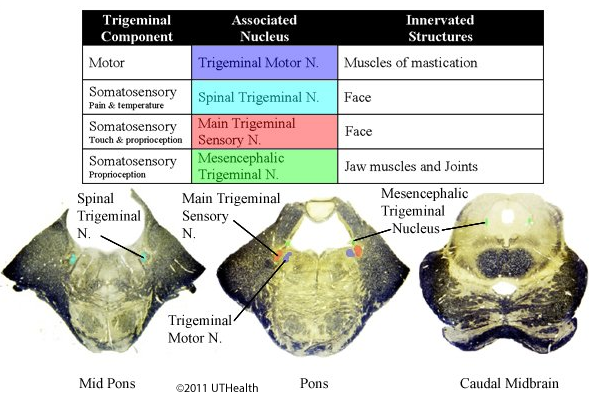
Components of the Trigeminal Nerve include
- Motor: The axons of the trigeminal motor nucleus exit at the lateral margin of the pons. The axons form the bulk of the mandibular division of the trigeminal nerve.
- Somatosensory: The trigeminal nerve fibers carrying crude touch, pain and temperature information from the face form the spinal trigeminal tract and terminate in the spinal trigeminal nucleus.
- Somatosensory: Trigeminal nerve fibers that carry information about discriminative touch and proprioception from the face end on the cells in the main sensory trigeminal nucleus.
- The neurons of the mesencephalic trigeminal nucleus are the cells of origin (first-order) of trigeminal nerve afferents innervating the jaw muscles and joints. They are concerned with proprioceptive sensibility of the jaw only and are involved in the controlling the force of bite.
 The trigeminal motor nucleus provides motor innervation to the muscles of mastication, the tensor tympani and the tensor veli palatini. Damage to the trigeminal motor neurons results in paralysis of the masticatory muscles. The force of bite is reduced in power, and the jaw deviates toward the injured side on opening the mouth. With bilateral damage, the lower jaw droops, but the mouth can be closed by means of the facial muscles. However, chewing is impossible and swallowing is difficult.
Among the inputs to the trigeminal motor nucleus are the collaterals from the mesencephalic trigeminal nucleus for reflex control of jaw muscles. Additional inputs from the other sensory trigeminal nuclei provide reflex control of jaw muscles to superficial stimuli, especially from the lingual and oral mucus membranes.
The corticofugal input to the trigeminal motor nucleus is both direct and indirect via the reticular formation and bilateral with a slight contralateral dominance. Central or upper motor neuron lesions lead to little disturbances unless they are bilateral, as in pseudobulbar palsy. In this disease, cerebral vascular disorders produce areas of necrosis, which interrupt the corticobulbar fibers. Volitional movements of the jaw are impaired and the jaw jerk reflex is usually increased.
The somatosensory components of the trigeminal nerve (facial skin, eyes, oral and nasal cavities and dura) and those of the vagus (outer ear canal, pinna, and dura), glossopharyngeal (outer ear canal, ear drum, and middle ear cavity) and facial (pinna) nerves end in the spinal trigeminal nucleus (crude touch, pain and temperature) and main sensory trigeminal nucleus(discriminative touch). The axons of the mesencephalic trigeminal nucleus travel in the trigeminal nerve to innervate and provide proprioceptive information from the jaw muscles and joints.
Cranial Nerve (V) Exam:
- Assess light touch sensitivity: Ask patient to respond when the examiner touches their skin with a piece of cotton/gauze on both sides of the forehead, followed by both sides of the cheeks, followed by both sides of the jaw. Ask patient if the sensation is the same on each side.
- Test muscles of mastication (temporal/masseter): Ask patient to bite down or clench teeth while the masseter or temporalis muscles are felt bilaterally.