Lab 9 (ƒ 10) - Cranial Nerve Nuclei and Brain Stem Circulation
Cranial Nerve VI-Abducens Nerve
The abducens (VI) nerve is motor in function and innervates the lateral rectus muscle of the eye. We have already encountered the abducens nucleus and nerve rootlets in the previous sections.
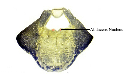
 The abducens nucleus lies within the caudal third of the pons in the facial colliculus. It contains both motor neurons and interneurons. The axons of its motor neurons form the abducens nerve and innervate the lateral rectus muscle of the eye. When the eye is directed straight ahead, contraction of the lateral rectus results in the external rotation or abduction of the eye. When the eye is elevated or depressed above or below the horizon, contraction of the lateral rectus will further elevate or depress the globe. The abducens interneurons send control signals to contralateral oculomotor neurons (via the medial longitudinal fasciculus) which control the medial rectus.
Lesions of the abducens nerve result in a paralysis of the ipsilateral lateral rectus muscle and double vision (diplopia). The affected eye is strongly adducted by the unopposed medial rectus muscle resulting in an internal strabismus. Recall that the abducens nucleus interneurons receive input from the pontine paramedian reticular formation (PPRF) and relays cortical influences that control conjugate horizontal eye movements. Consequently, damage to the abducens nucleus results in an inability to direct either eye towards the side of the lesion (lateral gaze paralysis).
The abducens nucleus receives inputs from vestibular nuclei via the medial longitudinal fasciculus and from the midbrain via the reticular formation, as well as internuclear inputs from the trochlear and oculomotor nuclei. The vestibular inputs are concerned with vestibular control of horizontal eye movements (i.e., vestibulo-ocular and smooth pursuit). Midbrain supranuclear inputs are involved in the control of voluntary and reflex lateral eye movements. The internuclear fibers from the other extraocular motor nuclei are involved in coordinating eye muscle action that enable conjugate and converging eye movements.
Cortical influences on abducens neurons are indirect and mediated by midbrain and pontine structures. Following a vascular accident, which damages the corticofugal fibers in the internal capsule, there may be a transient inability to look toward the side opposite the lesion on command (voluntary saccades), although reflex movements are well preserved.
Cranial Nerve (III, IV, VI) Exam: Extraocular Movements:
- Ask to look horizontally to the right
- Ask to look horizontally to the left
- Ask to look vertically up and down
- Assess tracking response: Hold patient's chin steady and ask patient to follow finger with their eyes, as examiner traces an "H" pattern about one meter from the patient's eye. From the middle, move finger about a foot to the right and pause; up and pause; down and pause; back to the midline; then cross midline and repeat sequence on the opposite side.
- Assess convergence response: Ask patient to look first at a distant point ahead, then at a target about five inches in front of the patient's nose.
