Lab 8 (ƒ7) - Visual System
Infranuclear Portion - Extraocular Muscles
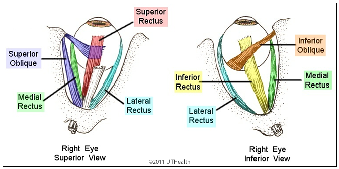 The individual movements of a single eye are known as ductions. For example, infraduction (looking down), abduction (looking laterally or outward), adduction (looking medially or inward toward the nose), and supraduction (looking up). Each eye is moved by six extraocular muscles: the medial and lateral rectus, the superior and inferior rectus, and the inferior and superior oblique muscles. Horizontal movements involve the coordinated actions of the lateral and medial rectus. For example, the lateral rectus contracts and the medial rectus relaxes to abduct the eye. The opposite actions must occur to adduct the eye. Vertical movements involve the coordinated actions of the superior and inferior rectus muscles. In a supraduction, the superior rectus contracts and inferior rectus relaxes. Oblique and torsional movements of an individual eye are produced by particular patterns of excitation and inhibition involving all the muscles.
Three cranial nerves innervate the extraocular muscles of a single eye as follows:
- Abducens (VI): lateral rectus (nerve fibers are ipsilateral to their cells of origin in the abducens nucleus)
- Oculomotor (III): the inferior rectus, medial rectus, and inferior oblique (nerve fibers are all ipsilateral to their cells of origin in the oculomotor nucleus) and the superior rectus (nerve fibers are contralateral to their cells of origin in the oculomotor nucleus)
- Trochlear(IV): the superior oblique (nerve fibers are contralateral to their cells of origin in the trochlear nucleus)