Lab 8 (ƒ7) - Visual System
Gross Anatomy of the Eye
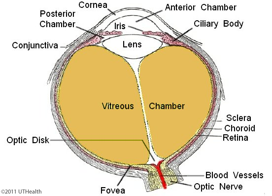 This is a horizontal section of the eye viewed from a superior position. The sclera is the connective tissue capsule, which appears as the andquot;whitesandquot; of the eyes. The cornea is the anterior, transparent portion of the outer coat. Fibers of the optic nerve pierce the sclera at the optic disc. Retinal blood vessels also enter and exit the eye at the optic disc. The choroid is a pigmented and richly vascularized membrane that lines the inner surface of the sclera. The iris extends from the choroid and rests on the anterior surface of the lens. You can see the ciliary body, which is interposed between the choroid and the iris. Note that the ciliary muscles are located in the base of the ciliary body. The suspensory ligaments attach the ciliary body to the lens capsule. The iris contains two sets of muscles (the sphincter and dilator muscles) that control the size of its opening, the pupil. The innermost coat of the eye, the retina, can be seen where it has separated from the choroid.
The anterior chamber of the eye is located between the cornea and lens and normally contains the fluid aqueous humor. The posterior chamber is located between the iris and anterior periphery of the lens and extends posteriorly behind the lens. The posterior chamber also contains the fluid aqueous humor. The vitreous chamber is the larger chamber located behind the lens. It normally contains a gelatinous mass called the vitreous humor or vitreous body.
The Fovea is part of the macula (a yellowish area lateral to the optic disc). The fovea is seen as a depression in the retina lateral to the optic disc. Weandrsquo;ll cover the fovea in the next section.