Lab 8 (ƒ7) - Visual System
Gross Anatomy of the Eye
LGN and LGB are used interchangeably and serve the same function. The 1° afferents of the visual system are the rods and cones of the retina (i.e., the photoreceptors) and the 2° afferents are the bipolar cells of the retina. The axons of the retinal ganglion cells (3° afferents) emerge from the eye to form the optic nerve. This andquot;nerveandquot;, which in humans contains approximately 1 million fibers, is actually a fiber tract of the CNS as embryologically the retina derives from the brain.
Within the cranial cavity, before entering the brain, the fibers from the nasal regions of the two retinas decussate to form the optic chiasm. Beyond the optic chiasm, the fibers from the temporal area of the ipsilateral retina join with the fibers from the nasal area of the contralateral retina to form the optic tract. 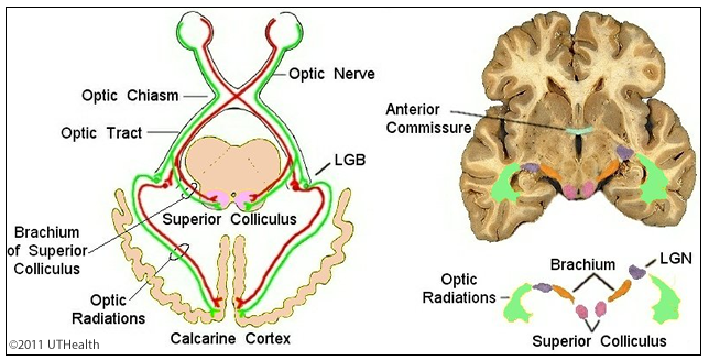 The fibers of the optic tract enter the brain at the level of the posterior diencephalon and may:
- Terminate in the lateral geniculate body (LGB) of the thalamus, or
- Pass on to the midbrain in the brachium of the superior colliculus to terminate in thesuperior colliculus (tectal area) and in pretectal areas.
The human LGB consists of six distinct layers: the cells in layers 1, 4 and 6 synapse with fibers from the contralateral eye (nasal retina) and cells in layers 2, 3 and 5 synapse with fibers from the ipsilateral eye (temporal retina). Thus, each cell in the LGB receives information from only one eye (monocular input). The axons of the LGB (4° afferent) travel in the optic radiations of the internal capsule to the primary cortical receiving area for the visual system, the calcarine cortex (also called the striate cortex) of the occipital lobe. The axons of the superior colliculus and pretectal area travel to a number of structures and are believed to play a role in reflex visual responses.
This figure is a diagram of these pathways. Decussating and non-decussating fibers are shown in red and green, respectively. On the right is a horizontal section at the level of the anterior commissure. The fibers of the optic tract enter the superior colliculus (purple) via the brachium of the superior colliculus (orange). Alternatively, the fibers may synapse in the lateral geniculate body onto fibers of the optic radiation.