Lab 8 (ƒ7) - Visual System
Microscopic Anatomy of the Eye
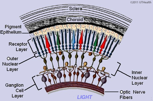 The sclera is the layer at the top of the figure. The interior of the eye, the vitreous chamber is on the bottom. Immediately internal to the sclera is the choroid, which is dark as it is highly pigmented. The retinal pigment epithelium appears as a thin dark layer that forms a boundary between the neural retina and the choroid.
The neural retina is formed by layers of neurons and glia. The light-sensitive portions (the outer segments) of the photoreceptors (the rods and cones) are located in the receptor layer that lies adjacent to the retinal pigment epithelium. The cell bodies of retinal neurons form the darkly stained retinal layers: the outer nuclear layer (photoreceptor soma), the inner nuclear layer (horizontal, bipolar and amacrine soma) and the ganglion cell layer (retinal ganglion soma).
Between the outer and inner nuclear layer, the area of synapse between the photoreceptor cells, horizontal cells and bipolar cells forms the outer plexiform layer. The inner plexiform layer lies between the inner nuclear layer and the ganglion cell layer and contains the area of synapses between the bipolar, amacrine and ganglion cells. Interior to the ganglion cell layer, the axons of the retinal ganglion cells form the optic nerve fiber layer in their course to the optic disc. These axons exit the eye at the disc and form the optic nerve, cranial nerve II.