Lab 2 (ƒ4) - External and Internal Anatomy of the Spinal Cord
Posterior Column Medial Lemniscal Pathway - Preview (continued)
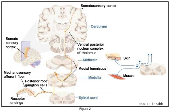
Take a look at these figures. Figure 1 is a series of microscopic cuts through the spinal cord, medulla, pons, midbrain, and diencephalon. Figure 2 is a diagrammatic representation of the posterior column-medial lemniscal pathway. Identify the structures associated with this pathway at each level. The text will provide a brief summary of each level. Try to refer to the text as you scroll though the levels. After you are finished with this section, we will go through each level in more detail and identify the necessary landmarks and associated structures.
Lumbar Spinal Cord
The 1° afferent fibers, which carry discriminative touch and proprioceptive information from the lower body, enter the cord via the medial division of the posterior root and join the fasciculus gracilis. The 1° afferents branch and have collaterals that synapse in deep laminae of the spinal gray matter, and long branches that travel upward in the posterior columns of the spinal cord without crossing. We will see later the levels at which they synapse and cross.
Cervical Spinal Cord
At and above the level of T6, the ascending central processes of the 1° afferents collect within the fasciculus cuneatus, which is lateral to the posterior intermediate sulcus. Fibers from the lower segments are still collected and ascend within the fasciculus gracilis. The fasciculus gracilis and fasciculus cuneatus collectively form the posterior funiculus, which is also called the posterior (dorsal) column.
 Caudal Medulla
This is a section through the caudal medulla, and includes the sensory decussation. Locate the fasciculi gracilis and cuneatus, which send their fibers into their respective nuclei. Notice that the nucleus gracilis is much smaller than the nucleus cuneatus. The axons of these two nuclei, 2° somatosensory afferents, sweep through the interior of the medulla as the internal arcuate fibers, form the sensory decussation and collect in the contralateral medial lemniscus.
Mid Pons
This section passes through the mid-pons. The fibers of the medial lemniscus continue to shift laterally to form a flat band of longitudinally coursing, crossed, 2° somatosensory afferent fibers near the anterior border of the pons tegmentum. Recall that the neurons of the posterior column-medial lemniscal pathway process information used for discriminative touch and proprioception from the body.
Rostral Midbrain (Transverse section)
This section passes through the rostral midbrain. In the midbrain, the crossed 2° somatosensory afferent fibers carrying discriminative touch and proprioception information from the body continue to ascend in the medial lemniscus. At this level of the midbrain, the fibers of the medial lemniscus have been displaced laterally and are beginning to move posteriorly along the lateral edge of the midbrain tegmentum.
Rostral Midbrain (Coronal Section)
This is a coronal section that passes through the rostral midbrain, caudal diencephalon and caudal basal ganglia. The midbrain structures include the crus cerebri, substantia nigra, and red nucleus. The third ventricle has replaced the cerebral aqueduct. Located posterior to the midbrain structures is the thalamus (part of the diencephalon.) Lateral to the midbrain and thalamus are the posterior limb of the internal capsule and the caudal remnants of the basal ganglia (i.e., the globus pallidus and putamen). The medial lemniscus extends from the lateral surface of the red nucleus, fans out posterolaterally towards the ventral posterolateral (VPL) nucleus of the thalamus where it terminates. The VPL neurons (3° somatosensory afferents) send their axons in the posterior limb of the internal capsule to the postcentral gyrus of the parietal lobe. Recall that the postcentral gyrus of the parietal lobe constitutes the primary cortical receiving area for the somatosensory systems.
What’s Next
With that overview, we will next go into more detail for each of the levels. More structures will be introduced in the following sections. You will relate these structures to the ones you have just learned, so make sure you know the structures in this section before moving on.