Lab 2 (ƒ4) - External and Internal Anatomy of the Spinal Cord
Spinal Cord Meninges - Introduction
The CSF and the spinal meninges protect the spinal cord within the vertebral column. These membranes are continuous with the cerebral meninges at the foramen magnum and the spinal subdural and subarachnoid spaces and are thus continuous with the corresponding cranial spaces. This is important since bleeding, infection, or inflammation in the cerebral subarachnoid space can be detected by sampling CSF from the lumbar subarachnoid space (i.e., using a lumbar puncture).

The spinal dura extends the entire length of the vertebral column down to the level of the second sacral vertebra (S2). The epidural space contains veins and fat and separates the dura from the periosteum of the vertebrae. Note that the nerve roots traverse this space, allowing effective introduction of local anesthetics into this space, particularly for obstetric purposes (as in an epidural block).
The anterior and posterior nerve roots penetrate the dura at the intervertebral foramen, and the sleeve of dura, which envelops these roots, becomes continuous with the epineurium of the peripheral nerves.
The arachnoid extends with the dura down to the level of the second sacral vertebra (S2). It is separated from the dura by the subdural space which is more potential than real. The subarachnoid space is wider and contains cerebrospinal fluid. Since the arachnoid extends to the second sacral vertebra, but the spinal cord ends at the level of the upper lumbar vertebra, a large cistern, the lumbar cistern, is formed below the second lumbar vertebra. CSF can be safely sampled without puncturing the spinal cord by introducing a needle into this lumbar cistern.
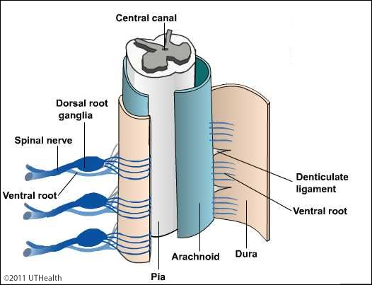
The pia adheres closely to the spinal cord. The blood vessels of the spinal cord lie on the surface of the pia. The denticulate ligaments are double folds of pia that extend from the lateral aspect of the cord to the inner surface of the dura, where they are attached on each side as a series of some 21 tooth-like extensions. These 21 denticulate ligaments are extensions of the pia that allow the cord to “float” in the spinal canal. The points of attachment of the dentate ligaments alternate with the points of exit of nerve roots. At the conus medullaris, the filum terminale, which is also derived from the pia, extends caudally to penetrate the dura and continue as the coccygeal ligament.