Lab 5 (ƒ8) - Higher Motor Function
Thalamic Nucleus
In the following sections, note the relationship of the thalamus to the basal ganglia on the horizontal and coronal slices. Be certain you can identify the various components of the basal ganglia and internal capsule on a series of horizontal and coronal slices. The caudate, putamen, globus pallidus and substantia nigra are extremely important physiologically in motor control. Huntington's chorea and Wilson's disease are examples of diseases that affect these structures. You will be unable to identify most of the thalamic nuclei in gross brain slices.
 Notice that throughout most of the thalamus the internal medullary lamina divides the thalamus into a medial group, consisting of the dorsomedial (DM) nucleus, and a lateral group, consisting of several thalamic nuclei. Anteriorly, the internal medullary lamina encloses the anterior nucleus, separating it from the ventral anterior (VA) nucleus. The VA nucleus only spans a small anterior to posterior distance. The anterior thalamic nucleus receives input from the mammillary bodies and projects to the cingulate gyrus. The anterior nucleus is part of the limbic system. The VA thalamic nucleus, the most rostral of the lateral thalamic group is one of the major thalamic motor nuclei.
As we move back the VL replaces the VA, so that the internal medullary lamina now separates the anterior nucleus from the ventral lateral nucleus. Like the VA, the VL nucleus is also a major thalamic motor nucleus. VL is often included with VA and called VA/VL, since both nuclei are involved in motor function. VA/VL receive input from the globus pallidus, substantia nigra and the cerebellar nuclei and sends output to frontal cortex, including motor cortex.
The DM nucleus receives input from limbic structures and sends its axons chiefly to the prefrontal cortex. The DM is part of the olfactory system.
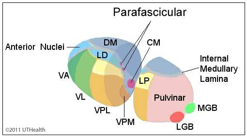
As we move further back the dorsomedial (DM) nucleus of the thalamus becomes more prominent. The lateral dorsal (LD) nucleus replaces the anterior nucleus in position, and the lateral group consists of the LD, lateral posterior (LP) nucleus, ventral posteromedial (VPM) and ventral posterolateral (VPL) nuclei. The centromedian (CM) nucleus is located within the internal medullary lamina and constitutes part of the intralaminar nuclear group along with the parafascicular nuclei. The LP nucleus receives inputs from the superior colliculus, other thalamic nuclei and from the parietal lobe and projects back to the parietal lobe. Its function is not well understood. The LD receives input from the cingulate gyrus and sends output back to the cingulate gyrus. The CM receives input from a number of noncortical sources (e.g., the reticular formation, spinal cord and globus pallidus) and sends its axons to the basal ganglia and to diffuse cortical areas. The CM is believed to be involved in motor function. The VPL receives the ascending fibers of the medial lemniscus and the spinothalamic tract. The VPL is the sensory thalamic nucleus of the somatosensory system. Recall that the VPM receives the fibers of the ventral trigeminal lemniscus.
As we move further back, the DM and LD are no longer present and the pulvinar is the most caudal part of the lateral thalamic group. Note the location of the geniculate bodies with respect to the pulvinar. The pulvinar receives input from several sensory systems, and sends axons to the parietal, temporal, and occipital cortices. As you already know, the medial geniculate body is the thalamic sensory nucleus for the auditory system and the lateral geniculate body is the thalamic sensory nucleus for the visual system.
When viewing coronal sections through the thalamus, keep the following rules of thumb in mind:
- When you can see A and there is no DM, the lateral group consists of VA.
- When you can see DM, but not CM or VPM, the lateral group consists of the VL.
- When you can see DM, CM and VPM, LD replaces A and the lateral thalamic group consists of the LD, LP, VPM and VPL.