Lab 4 (ƒ3) - The Ventricles and Blood Supply
Cranial Nerves of the Pons
Part of the posterior surface of the pons forms the rostral half of the floor of the fourth ventricle. The stria medullaris in the floor of the fourth ventricle forms the caudal border of the pons.
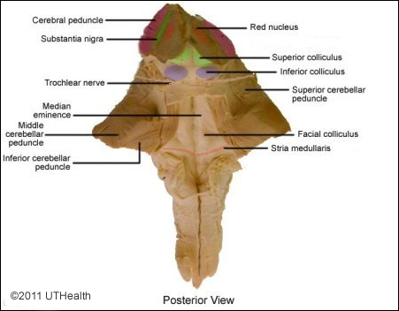 Rostral to the stria medullaris, the paired elevations bordering the median sulcus are called the facial colliculi. The elevation which continues rostral to the facial colliculus is known as the median eminence. A shallow groove, the sulcus limitans extends from the pons into the medulla and separates the facial colliculus and medial eminence from the more laterally located sensory or vestibular area. The superior cerebellar peduncles appear on the rostral end of the posterior pons as two bands of fibers running in the walls of the fourth ventricle.
The anterior surface of the pons is characterized by a massive band of transverse bundles of nerve fibers that converge posteriorly on each side to form the middle cerebellar peduncles. Notice the shallow groove that is running along the anterior midline. This is the pontomedullary sulcus and it normally contains the basilar artery.
There is one cranial nerve associated with the pons proper, the trigeminal nerve (cranial nerve V). Three other cranial nerves are located at the pontomedullary junction: the abducens nerve (cranial nerve VI), the facial nerve(cranial nerve VII), and the vestibulocochlear nerve (cranial nerve VIII).
Find the trigeminal nerve as it leaves the brain stem from the anterolateral aspect of the pons about halfway between its rostral and caudal borders. The trigeminal nerve is made up of two bundles, the larger portio major, which is sensory to the face, and a smaller portio minor, which is motor to the muscles of mastication.
Observe the abducens nerve root fibers as they leave the brain stem in the pontomedullary sulcus a little lateral to the pyramid. Abducens nerve fibers innervate the ipsilateral lateral rectus of the eye. A lesion of the right abducens nerve would result in failure to abduct the denervated eye.
Find the facial nerve and its nervus intermedius, which emerge at the caudal border of the pons lateral to the abducens nerve. The facial nerve contains, among other things, motor axons innervating the muscles of facial expression. The nervus intermedius consists predominantly of sensory axons innervating taste receptors on the anterior two thirds of the tongue.
Locate the vestibulocochlear nerve as it emerges caudal to the pons and lateral to the facial nerve. The facial and vestibulocochlear nerves converge further along their course towards the periphery in the internal auditory meatus. The vestibular portion of cranial nerve VIII carries information about the position and movement of the head relative to earth (i.e., gravity) as determined by vestibular receptor cells in the inner ear. The cochlear portion of this nerve carries auditory information encoded by receptors in the cochlea of the inner ear. The auditory receiving area in the cerebral cortex is the superior bank of the superior temporal gyrus.
The region where cranial nerves VII, VIII and IX are found is called the cerebellopontine or cerebellomedullary angle. Tumors can develop in that region and produce symptoms involving all the nerves within the angle.