Lab 4 (ƒ3) - The Ventricles and Blood Supply
Cranial Nerves of the Midbrain
The brain stem consists of the medulla, pons and midbrain. Most of the cranial nerves (III to XII) take origin or terminate in the brain stem.
A. The Midbrain or Mesencephalon
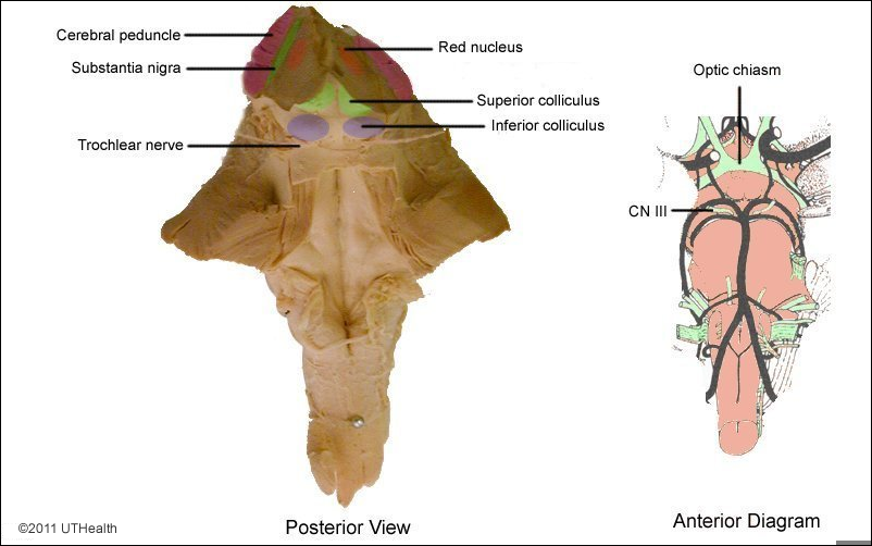
In the posterior view examine the cut edge of the midbrain and look for two black bands posterior to the two cerebral peduncles. These lines are the substantia nigra. Locate the two large reddish-gray structures, the red nuclei, located along the midline just posterior to the substantia nigra. The colors of these two midbrain nuclei in the unstained gross specimens result from pigment in their neurons. Observe also the small channel formed by the cerebral aqueduct at the midline. As you learned in Exercise 1, there are four small, round elevations, the inferior colliculi and the superior colliculi, which form the tectum or roof of the midbrain.
The primary function of the Superior colliculi is for Sight, and the primary function of the inferior colliculi is auditory.
On the anterior surface of the midbrain two large longitudinal fiber bundles, the cerebral peduncles are separated by a deep groove called the interpeduncular fossa.
There are two cranial nerves associated with the midbrain, the oculomotor (cranial nerve III) and the trochlear (cranial nerve IV). Find the oculomotor nerve root as it emerges from the anterior aspect of the midbrain - medial to the crus cerebri, in the interpeduncular fossa. Motor fibers in the nerve innervate the intrinsic muscles of the eye and all the extrinsic muscles of the eye except for the lateral rectus and the superior oblique. Find the trochlear nerve as it leaves the midbrain from the posterior surface, just caudal to the inferior colliculus. This is the only cranial nerve to leave the brain stem posteriorly. Axons of the trochlear nerve innervate the superior oblique muscle of the eye.