Lab 4 (ƒ3) - The Ventricles and Blood Supply
Vertebral-Basilar System
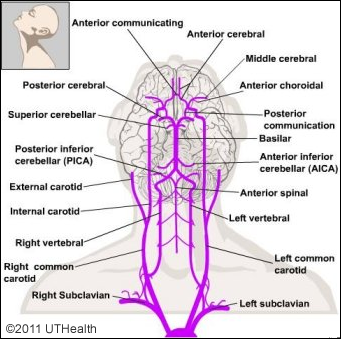
Except for the most rostral portion of the crus cerebri, the entire blood supply of the brain stem (i.e., midbrain, pons and medulla) and cerebellum is derived from the vertebral-basal system. As you become more familiar with the functional anatomy of the brain stem, return to this figure and consider the consequences of occlusion of the various vessels.
 1. Vertebral Arteries
The paired vertebral arteries pass over the anterior surface of the medulla and give rise to the posterior spinal arteries, the anterior spinal arteries and the posterior inferior cerebellar arteries. The paired vertebral arteries pass rostrally and unite near the pontomedullary junction to form the basilar artery.
2. Posterior Spinal Arteries
As noted above, the posterior spinal arteries arise from the vertebral arteries and descend along the posterolateral aspect of the medulla and spinal cord. Follow the posterior spinal artery as it passes posteriorly along the medulla. Branches of this artery supply posterior areas of the medulla that are also supplied by branches of the vertebral artery (caudally) and the posterior inferior cerebellar artery (rostrally). Note that the posterior spinal arteries descend to, and supply the spinal cord.
3. Anterior Spinal Arteries
See if you can find branches of the anterior spinal arteries which supply the anterior and midline regions of the medulla. These arteries merge to form a single artery which descends in the anterior median fissure of the spinal cord.
4. Posterior Inferior Cerebellar Arteries
The posterior inferior cerebellar artery usually arises from the vertebral artery, but its position of origin is quite variable on the parent vessel. On the whole brain follow the posterior inferior cerebellar artery (PICA) as it travels along the lateral and posterior surface of the medulla. In its course along the medulla it gives off small branches to supply posterolateral regions of the medulla. Follow this artery as it courses up to the cerebellum to supply the inferior surface of the cerebellum and the choroid plexus of the fourth ventricle.
5. Basilar Artery
The basilar artery is a large midline arterial trunk formed by the fusion of the vertebral arteries on the anterior surface of the brain stem. Locate the basilar artery on the whole brain specimen. In some specimens the anterior inferior cerebellar artery may come off the basilar artery near its origin. The superior cerebellar artery is derived from the basilar artery near the pons-midbrain junction. In most specimens the basilar artery branches caudal to the mammillary bodies to form the posterior cerebral arteries.
6. Anterior Inferior Cerebellar Arteries
The anterior inferior cerebellar arteries arise from the basilar artery near the pontomedullary junction. It is the most caudal large artery originating directly from the basilar artery. Locate this branch of the basilar artery and follow its course to the cerebellum. It supplies inferior parts of the cerebellum lateral to that supplied by the posterior inferior cerebellar artery. It also provides branches to the caudal pontine tegmentum and to the choroid plexus of the fourth ventricle.
7. Paramedian and Circumferential Branches
Along its rostral course, the basilar artery gives off paramedian branches that enter the brain stem on its anterior surface near the midline. If you gently lift up the basilar artery, you will observe these small delicate branches penetrating the brain stem. These branches provide blood to structures in the basal and medial tegmental regions of the brain stem. The circumferential branches arise from the basilar artery and pass around and the brain stem enter along its anterolateral and posterolateral surfaces. See if you can find a long circumferential branch and follow it to the posterolateral surface of the brain stem.
8. Superior Cerebellar Arteries
The superior cerebellar arteries arise from the basilar artery near the pons-midbrain junction. Follow the artery as it travels along the lateral surface of the pons and note that it subdivides to provide branches to the superior surface of the cerebellum and to the deep cerebellar nuclei and white matter. Small branches also provide blood to the rostral part of the posterior pontine tegmentum.
9. Posterior Cerebral Arteries
The posterior cerebral arteries are usually formed by the rostral bifurcation of the basilar artery. The posterior cerebral artery provides cortical branches that supply the cuneus and lingual gyri of the occipital lobe, the inferior surface of the temporal lobe and the lateral occipital gyri. Because it provides the major blood supply to the visual cortex, occlusion of this artery results in visual field defects, contralateral to the site of injury.