Lab 4 (ƒ3) - The Ventricles and Blood Supply
Cranial Nerves of the Medulla
The anterior median sulcus along the midline of the anterior medulla divides two longitudinal eminences called the pyramids. This sulcus is obscured by small fiber bundles in the pyramidal decussation near the junction of the medulla and the spinal cord. Rostrally, the preolivary sulcus, containing rootlets of the hypoglossal nerve (cranial nerve XII) divides the pyramids from the olivary eminence. The postolivary sulcus borders the olivary eminence laterally and contains the rootlets of the glossopharyngeal nerve (cranial nerve IX), the vagus nerve (cranial nerve X), and the spinal accessory nerve (cranial nerve XI).
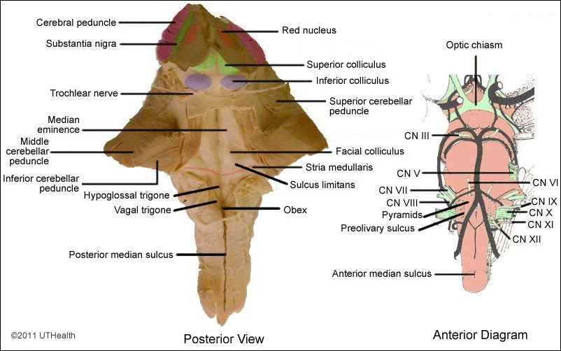
In posterior view, the medulla is seen to consist of a closed, caudal portion that contains a continuation of the spinal cord central canal and an open, rostral portion that forms part of the floor of the fourth ventricle. The apex of the V-shaped caudal border of the fourth ventricle is known as the obex. The posterior median sulcus extends rostrally from the spinal cord to the obex. Lateral to this sulcus is a longitudinal bulge, the gracilis tubercle or clava. At a level slightly rostral to the clava, the cuneate tubercle appears as a small bulge laterally. The tuberculum cinereum is a slight swelling located lateral to the cuneate tubercle. The tuberculum cinereum becomes displaced laterally in the rostral (open) medulla by the large inferior cerebellar peduncles or restiform body.
The stria medullaris in the floor of the fourth ventricle marks the rostral border of the medulla. The median sulcus divides the floor of the ventricle along the midline, while the sulcus limitans divides each half of the medulla into medial (motor) areas and lateral (sensory) areas. The motor area of the medulla consists of two paired elevations called trigones. The paired elevations nearest the midline are called the hypoglossal trigones. The vagal trigones are located just lateral to the hypoglossal trigones. Lateral to the sulcus limitans is the sensory or vestibular area. The inferior cerebellar peduncles appear as two ridges extending along the sides of the ventricle on the lateral surface of the medulla.
Find the glossopharyngeal nerve, which appears as a series of rootlets that emerges from the brain stem in the postolivary sulcus. This nerve innervates the posterior third of the tongue, other areas of the oral cavity, part of the ear and parasympathetic (otic) ganglion. Caudal to the rootlets of the glossopharyngeal nerve are the rootlets of the vagus nerve that also emerge from the brain stem at the postolivary sulcus. The nerve fibers of the vagus nerve innervate the oral and ear cavities, the larynx and pharynx, and parasympathetic ganglia of trunk and abdominal viscera. The spinal accessory nerve (XI) is formed by the union of rootlets emerging from the postolivary sulcus caudal to the vagus roots with rootlets from upper cervical levels of the spinal cord. The accessory nerve innervates the ipsilateral sternocleidomastoid and trapezius muscles.
The hypoglossal nerve (XII) rootlets emerge from the preolivary sulcus just lateral to the pyramids. The fibers of the hypoglossal nerve innervate the somatic skeletal muscles of the tongue.