Lab 4 (ƒ3*) - The Ventricles and Blood Supply
The Ventricles
The ventricular system is an interconnected series of spaces within the brain, containing cerebrospinal fluid (CSF) and the choroid plexus which produces the CSF. Your main concern at this time should be to acquire an appreciation of the overall shape and position of the ventricles within the brain. The specific neural structures which make up their walls will be studied in more detail in later exercises.
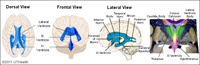
A. Lateral Ventricles
There are two lateral ventricles, one per cerebral hemisphere. The five subdivisions of the lateral ventricles include the anterior (frontal) horn, the body, the collateral trigone or atrium, the inferior (temporal) horn and the posterior (occipital) horn. In the hemisected brain with intact septum pellucidum, you will not be able to see the frontal horn, temporal horn, atrium, or the occipital horn of the lateral ventricle. However, be aware that as their names imply the frontal horn is located rostrally in the frontal lobe, the temporal horn inferiorly in the temporal lobe, and the occipital horn caudally in the occipital lobe. The atrium is the region of the lateral horn where the temporal and occipital horns are confluent with the body of the lateral ventricle. Rostrally the bodies of the lateral ventricles lie side-by-side, separated in part by the septum pellucidum.
Note that the superior surface of the thalamus forms part of the floor, the caudate body forms part of the lateral wall, and the inferior surface of the corpus callosum forms the roof of the lateral ventricle body. The choroid plexus may still be present in the exposed part of the lateral ventricle. Locate the interventricular foramen of Monro. Note that the lateral ventricles communicate with the third ventricle via these paired interventricular foramina. Choroid plexus is found in the lateral ventricle body, atrium, temporal horn and interventricular foramen.
B. Third Ventricle
The third ventricle is a thin, elongated cavity lying in the midsagittal plane of the diencephalon. The third ventricle contains choroid plexus which extends from the body of the lateral ventricle through the interventricular foramina. The lateral ventricle is connected with the third ventricle via the foramen of monro. The third ventricle is connected to the fourth ventricle via the cerebral aqueduct (also called the aqueduct of Sylvius).
The lateral walls of the third ventricle are formed by the medial aspects of the thalamus and the hypothalamus and its rostral wall is formed by the lamina terminalis.
C. Cerebral Aqueduct
The cerebral aqueduct connects the third ventricle to the fourth. Within the lateral ventricles, choroid plexus is found in the body, atrium, temporal horn and interventricular foramen.
Notice that the cerebral aqueduct is located immediately anterior to the midbrain tectum. The aqueduct is a midline tube-like structure a few centimeters in length and about as thick as a pencil lead. It connects the third ventricle to the fourth.
D. Fourth Ventricle
The fourth ventricle is shaped like an elongated pyramid, with its apex extending up into the cerebellum and its diamond-shaped base on the posterior surface of the pons and medulla. The fourth ventricle also contains choroid plexus which produces CSF. The CSF drains from the fourth ventricle into an enlargement of the subarachnoid space called the cisterna magna. There are three openings that lead from the fourth ventricle into the cisterna magna: the midline foramen of Magendie lies at the caudal end of the fourth ventricle and the two laterally situated foramina of Luschka are found at approximately mid-pontine level. These laterally situated foramina often can be identified on the inferior aspect of the brain by finding protrusions of choroid plexus in the subarachnoid space near the cerebellopontomedullary junction.
E. CSF Formation and Circulation
Cerebrospinal fluid is produced by modified capillaries called choroid plexus. These structures are located in the lateral, third and fourth ventricles. After formation in the ventricles, CSF passes into the cisterna magna via the foramina of Magendie and Luschka, circulates up and around the brain in the subarachnoid space, and is passively absorbed into the venous system via the arachnoid villi. The CSF of the ventricles communicates with the subarachnoid space only through the three foramina of the fourth ventricle. They are the foramina of Magendie and Luschka.