Lab 6 (ƒ9) Descending Pathways to the Spinal Cord
The Corticospinal Pathway
The figure summarizes the course of the corticospinal pathway from the motor cortex down to the spinal cord. Please read the following text and follow the pathway down before proceeding to the next section.
As we follow the corticospinal pathway down, we will also consider other corticofugal fibers. The corticofugal fibers include the corticorubral, corticopontine and corticobulbar (bulb generally refers to the medulla. Corticobulbar: from the cortex to the medulla) fibers in addition to the corticospinal fibers.
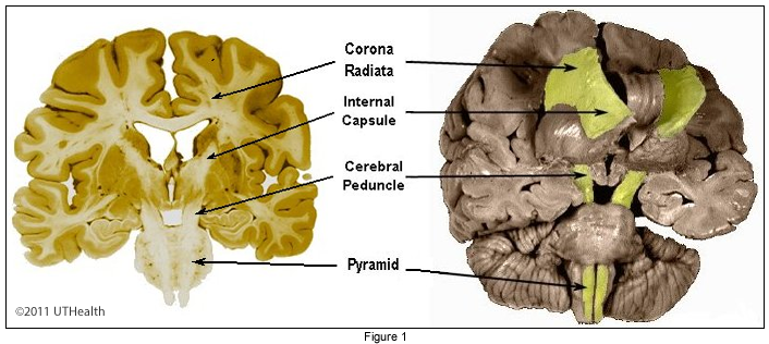
The fibers of the neurons in the precentral gyrus (motor cortex), frontal lobe, and parietal lobe enter the corona radiata, a radiating mass of white matter converging toward the internal capsule. Included in the internal capsule are other descending and ascending fibers.
The descending corticofugal fibers travel in the posterior limb of the internal capsule to the crus cerebri (AKA cerebral peduncle) of the midbrain. The fibers of the corticospinal tract are somatotopically organized: those fibers concerned with the Lower extremity are located Laterally while those concerned with the upper extremity and head are located more medially.
At the pontine level, corticopontine fibers (from the cortex to the pons) terminate in the pontine nuclei and pontine tegmentum. From here fibers travel to the cerebellum. The cerebral cortex influences the cerebellum via this corticopontocerebellar pathway.
At the base of the medulla the corticospinal fibers, along with the corticobulbar fibers form two large compact bundles, the pyramids. Corticobulbar fibers leave the pyramids along the entire course of the medulla to terminate in the medullary reticular formation and in certain cranial nerve nuclei.
Near the spinal cord most of the corticospinal fibers cross the midline in the pyramidal decussation and shift posteriorly. In the spinal cord, the corticospinal fibers of the pyramids form two tracts: (1) the crossed lateral corticospinal tract, which is located in the lateral funiculus, medial to the posterior spinocerebellar tract; and (2) the uncrossed anterior corticospinal tract, which is located in the anterior funiculus.
Most fibers of the lateral corticospinal tract terminate on interneurons, which influence motor neurons innervating the distal muscles of the hands and fingers, chiefly, and the proximal muscles.
Most of the anterior corticospinal fibers cross in the spinal cord anterior white commissure to terminate in the contralateral cord. Some remain uncrossed. The crossed fibers of the anterior corticospinal tract appear to terminate on interneurons. Those that do not cross terminate on neurons that influence the proximal limb muscles.
Remember that the majority of corticospinal fibers do not terminate directly upon the anterior horn motor neurons.
In the next section we will review cross sections along the course of the corticospinal pathway.