Lab 4 (ƒ3) - The Ventricles and Blood Supply
Superficial Blood Vessels - Internal Carotid System
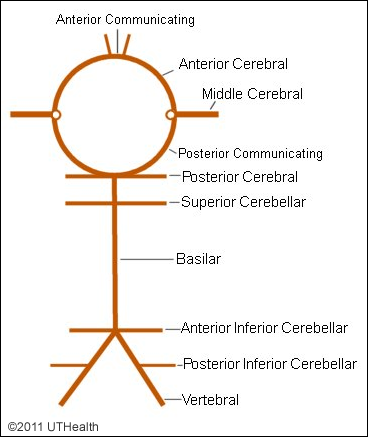 The brain is supplied by four large arteries: two internal carotid arteries and two vertebral arteries. You should recall that the internal carotid artery is a branch of the common carotid artery and the vertebral artery a branch of the subclavian artery. The internal carotid artery divides into the anterior and middle cerebral arteries. The paired vertebral arteries fuse at the caudal border of the pons to form the basilar artery.
A. Internal Carotid System
Inspect the base of the whole brain and identify all components of the circle of Willis, an anastomotic arterial ring that constitutes the major, but not exclusive, component of the intracranial collateral circulation. Variations in patterns of the circle of Willis are fairly frequent (Only about 50% of patients have a completely symmetrical circle of Willis). In various cerebrovascular diseases, especially arterial occlusion, an asymmetry may reduce the effective collateral circulation in parts of the cerebral hemisphere.
Locate the intracranial portion of the internal carotid on each side. Note its proximity to the optic nerve and optic chiasm. The anterior choroidal arteries should be identified as arising from the internal carotid artery lateral to the origin of the posterior communicating arteries. Besides supplying the choroid plexus of the lateral ventricles, the anterior choroidal arteries also provide branches to the core of the parahippocampal gyrus and the posterior portion of the internal capsule.
1. Anterior Cerebral Artery
Locate the anterior cerebral artery arising from the internal carotid body of each side. The major branches of the anterior cerebral artery supply the medial surface of the frontal and parietal cortex and much of the corpus callosum. Arterial border zones or leptomeningeal anastomoses exist between the anterior cerebral and middle cerebral arteries. They permit blood flow from cortical branches of one cortical arterial territory to the other. Small branches leave the anterior cerebral arteries and enter the anterior perforated substance to supply portions of the hypothalamus. The medial striate artery (Recurrent Artery of Heubner) arises from the anterior cerebral artery before the latter passes into the longitudinal cerebral fissure. The medial striate artery passes laterally and enters the brain to supply medial areas of the corpus striatum of the basal ganglia and anterior internal capsule.
2. Anterior Communicating Artery
The anterior cerebral artery of each side is connected to its partner by the anterior communicating artery, an important link in the circle of Willis. Identify the anterior communicating artery; it may be a single vessel, and may be large or small or hair-like. The anterior communicating artery permits shunting of blood from one anterior cerebral artery to the opposite side
3. Middle Cerebral Artery
Locate the main stem of the middle cerebral artery which gives off numerous penetrating branches, known as the lateral striate arteries or lenticulostriate arteries. These deep vessels supply most of the lateral areas of the corpus striatum of the basal ganglia and the internal capsule. If the lateral striate arteries are occluded the basal ganglia may be affected, causing motor deficits. The cortical branches of the middle cerebral artery are of great importance clinically as they supply most of the lateral aspect and some of the inferior aspect of the cerebrum.
4. Posterior Communicating Arteries
The posterior communicating arteries arise from the internal carotid arteries (in most cases), connect the latter to the posterior cerebral arteries, and are part of the circle of Willis. The posterior communicating arteries are often of unequal size. Small hair-like branches from the posterior communicating arteries supply the rostral and medial regions of the diencephalon.
5. Posterior Cerebral Arteries
The posterior cerebral arteries usually arise from the basilar artery but may originate from the internal carotids, another variation in pattern. The cortical branches of the posterior cerebral arteries supply the inferior part of the temporal lobe and the entire occipital lobe. The posterior choroidal artery is derived from the posterior cerebral artery and supplies the choroid plexus of the lateral and third ventricles and the superior aspect of the thalamus and the midbrain tectum.
NOTE that the internal carotid system supplies blood to most of the forebrain (e.g., the cerebral cortex, corpus callosum, basal ganglia and diencephalon). Thrombi passing through the internal carotid artery or any other vascular disease or injury associated with the internal carotid system may result in a devastating loss of function (stroke) or death.