Lab 3 (ƒ5) - Somatosensory, Viscerosensory and Spinocerebellar Pathways
Spinothalamic Pathways
There are two well-defined spinothalamic pathways, chiefly concerned with pain and temperature sensations and with crude touch. The “fast” conducting neospinothalamic pathway is involved in conveying the “sharp/cutting” pain elicited at the time tissue is damaged. 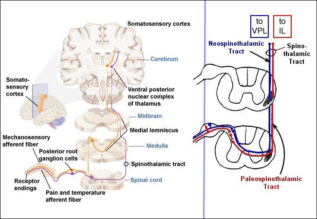 The “slower” conducting paleospinothalamic pathway is involved in conveying the “dull/burning” pain that accompanies the later inflammatory reaction in the damaged tissue as well as temperature and crude touch information. The archeospinothalamic pathway is a poorly defined pathway involved in a generalized sense of discomfort and diffuse pain.
The 1° afferents of the spinothalamic pathways send A-delta (neospinothalamic) and C (paleospinothalamic) fibers to the periphery where they form free nerve endings in skin, muscle, tendon, joint capsules and viscera. The central processes of these 1° afferent neurons are the pseudounipolar cells of the posterior root ganglia and enter the spinal cord in the lateral division of the posterior root. Recall that fibers entering via the medial division of the posterior root are concerned with tactile, pressure, proprioception, and vibration sensations. The lateral division axons branch and send fibers to the gray matter at the segment of entry and into the tract of Lissauer. The 1° afferents of the spinothalamic systems may end in the segment of root entry or one or two segments up. The neospinothalamics end on 2° afferents in the nucleus posteromarginalis. The paleospinothalamics end on 2° afferents in the substantia gelatinosa.
Neospinothalamic Pathway
The 2° neospinothalamic afferents (nucleus posteromarginalis axons) cross in the anterior white commissure to collect in the spinothalamic tract within the contralateral anterior and lateral (predominantly) funiculi. These 2° neospinothalamic afferent axons ascend the spinal cord and brain stem in the spinothalamic tract to terminate on 3° afferents in the ventral posterolateral (VPL) nucleus of the thalamus. Note that these 3° neospinothalamic VPL afferents differ from the VPL neurons synapsing with 2° afferent axons of the medial lemniscus. The 3° neospinothalamic VPL neurons send their axons to the primary somatosensory cortex (i.e., the postcentral gyrus of the parietal lobe).
Paleospinothalamic Pathway
The 2° paleospinothalamic afferents (axons of the substantia gelatinosa) travel a short distance to terminate in or near the ipsilateral nucleus proprius on 3° afferents. The axons of most paleospinothalamic 3° (nucleus proprius) afferents cross in the anterior white commissure to collect bilaterally in the spinothalamic tracts of the anterior (predominantly) and lateral funiculi. Along their ascending course to the thalamus, many of the paleospinothalamic 3° (nucleus proprius) afferents leave the spinothalamic tracts to terminate in the brain stem reticular formation or midbrain periaqueductal gray. The remaining paleospinothalamic 3° afferents remain in the spinothalamic tracts and ascend to terminate on 4° paleospinothalamic afferents in the intralaminar nuclei of the thalamus (IL in the figure). The projections of the intralaminar nuclei axons (4° paleospinothalamic afferents) are to diffuse areas of the cerebral cortex which are believed to play a role in poorly localized sense of pain. For example, destruction of the primary somatosensory cortex (i.e., the postcentral gyrus of the parietal lobe) does not seem as detrimental to the appreciation of painful stimuli as it is to the appreciation of other somatic sensations.
In the next section we will follow the spinothalamic pathway from the spinal cord to the cerebral cortex. Along the way, you will see another ascending pathway, the spinoreticular tract, which travels in close association with the spinothalamic tracts.