Lab 11 - The Limbic System
Review (continued)
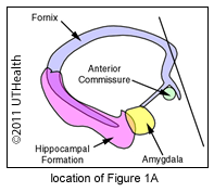 Figure 1, Layer A: This is the most rostral extension of the fornix (at the septal nuclei.) This section was cut at an angle; rostral to the anterior commissure on the left side and at the rostral edge of the transverse portion of the anterior commissure on the right side. You can see the fibers of the anterior commissure under the globus pallidus in the right half of the section. Remember that at the rostral level where the fornix approaches the anterior commissure, some fibers split off from the fornix and run anterior to the commissure as the precommissural fornix. The dark vertical bands in the septum pellucidum are fibers of the precommissural fornix. Most of these fibers originate in the hippocampus of the hippocampal formation, and are destined for the septal nuclei. The septal nuclei are located near the base of the septum pellucidum, rostral and superior to the anterior commissure.
The stria terminalis cannot be seen as it is terminating in the septal nuclei. The septal nuclei send their axons to a number of sites: (1) the hippocampal formation through the fornix, (2) the amygdala through the stria terminalis (3) the habenula through the stria medullaris thalami, and (4) the hypothalamus through the medial forebrain bundle.
On the right hand side of the image in the region inferior to the transverse segment of the anterior commissure is a structure called the substantia innominata. For a so-called un-named substance, it sure has a lot of names. It is also known as the basal nucleus of Meynert and nucleus basalis. It contains neurons that are basophilic and cholinergic and receives input from the limbic system. It is of interest because neurons in it selectively degenerate in Alzheimer's disease.
NOTE: The nucleus accumbens septi is not part of the septal nuclei. It is a basal ganglia structure that receives input from the hippocampal formation and amygdala, among other sources and projects to an extension of the globus pallidus (ventral pallidum). It is part of a dopaminergic pathway that has been implicated in the reinforcing effects of dependent drugs (e.g., opiates, cocaine and ethanol). Locate the olfactory tract (left) and the lateral olfactory stria (right). Notice that the caudate 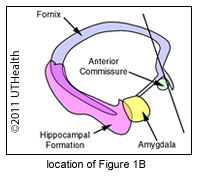 head appears to extend down to the superior surface of the anterior commissure.
Figure 1, Layer B: We are moving caudally following the fornix and stria terminalis as they travel away from the septal area. The anterior commissure is crossing the midline and appears as a dark band of transverse fibers passing below the globus pallidus. The basal nucleus of Meynert can be seen bilaterally inferior to the anterior commissure. The fornix column can be seen passing from the fornix body to the superior surface of the anterior commissure.
The stria terminalis is passing inferiorly, medial to the caudate, on its course to the hypothalamus and septal nuclei. The fibers of the stria terminalis originate in the amygdala. Inferiorly near the midline, the nuclei of the anterior hypothalamus are receiving precommissural fornix fibers. These fibers have looped around in front of the anterior commissure and traveled caudally to terminate in regions of the 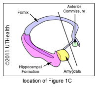 hypothalamus rostral and superior to the optic chiasm.
Figure 1, Layer C: The thick fiber bundle medial to the internal capsule genu is the postcommissural fornix, so called because they travel caudal to the anterior commissure. The fibers that constitute the postcommissural fornix arise mainly from the subiculum of the hippocampal formation. The majority of the fibers in the postcommissural fornix terminate in the mammillary body and thalamus. Locate the body of the right fornix.
Figure 1, Layer D: Anteriorly, the internal medullary lamina encloses the anterior (A) nucleus, separating it from the ventral anterior (VA) nucleus. The anterior thalamic nucleus receives input from the mammillary bodies and projects to the cingulate gyrus.
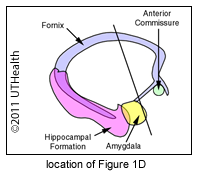 Following the course of the postcommissural fornix we see it passing inferiorly and caudally to terminate in the thalamus and posterior hypothalamus. The anterior (A) thalamic nucleus receives fibers from the postcommissural fornix and the mammillothalamic tract. The postcommissural fornix fibers that end in the anterior thalamic nuclei originate in the subiculum of the hippocampal formation. The precommissural fornix is a mixed system containing afferent fibers from the septal nuclei to the hippocampal formation and efferent fibers from the hippocampus. The anterior thalamic nucleus gives off thalamocortical projection fibers, which pass via the anterior limb of the internal capsule to the cingulate gyrus.
Locate the stria medullaris thalami. Its fibers originate in the septal nuclei and terminate in the habenula. The stria terminalis is located in the floor of the lateral ventricle body and is wedged between the caudate body and the thalamus (VA). The fibers of the stria terminalis originate in the amygdala and terminate in the septal nuclei and hypothalamus.
 Notice the postcommissural fornix passing inferiorly and caudally in its course to the mammillary bodies. Notice also the amygdala, anterior commissure, lentiform nuclei, claustrum, external capsule and extreme capsule.
Figure 1, Layer E: We are continuing in a caudal direction and are now at the level of the mammillary bodies of the hypothalamus. Small fascicles of postcommissural fornix fibers enter along the lateral margin of the mammillary body. The mammillothalamic tract is more prominent in this section and is situated superior to the mammillary body. It enters the mammillary body along its medial margin. The fornix is both afferent and efferent with respect to the mammillary bodies providing a bi-directional connection with the subiculum of the hippocampal formation. The mammillothalamic tract provides a bi-directional pathway connecting the mammillary bodies with the anterior thalamic nucleus.
Figure 1, Layer F: We are still moving caudally and the hippocampal formation has replaced the amygdala in the temporal lobe. Remember that the collateral sulcus separates the occipitotemporal gyrus from the parahippocampal gyrus. The parahippocampal gyrus extends from the collateral sulcus to the hippocampal sulcus. The parahippocampal gyrus consists of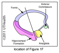 the entorhinal area. The subiculum is the transition zone between the hippocampus and parahippocampal gyrus. The hippocampus (also called Ammon's horn) and the dentate gyrus are located lateral and superior to the subiculum. The subiculum, hippocampus and dentate gyrus are collectively called the hippocampal formation. You can identify the dentate gyrus by the thin dark band of cells that make up its granule cell layer. A major afferent pathway into the hippocampal formation is via neurons in the entorhinal area. The entorhinal area receives axons from the piriform cortex, cingulate gyrus and amygdala and "relays" these inputs to the hippocampal formation. The septal nuclei and hypothalamus also send fibers back to the hippocampal formation via the fornix. However, remember that the fornix is the major efferent tract of the hippocampal formation. Identify the alveus and fimbria.
You should now realize that you can see the fornix in two places; the fornix fimbria near the hippocampal formation in the temporal horn and the fornix body in the lateral ventricle body. The stria terminalis is also visible in two places, in the roof of the temporal horn and in the floor of the lateral ventricle body. The dorsomedial (DM) thalamic nucleus receives amygdaloid fibers traveling in the diffuse ventral amygdalofugal pathway and sends fibers to the amygdala in the same pathway.