Lab 7 (ƒ6) - Auditory, Vestibular, Gustatory and Olfaction Systems
The Vestibular System - Introduction
The vestibular system is a proprioceptive system involved in the detection of head position and movement with respect to gravity. It functions to help maintain muscle tone and posture of the body, neck, head and eyes. The vestibular receptor cells are distributed in five discrete patches; i.e., in each crista of each semicircular duct and in the macula of the saccule and the macula of the utricle.
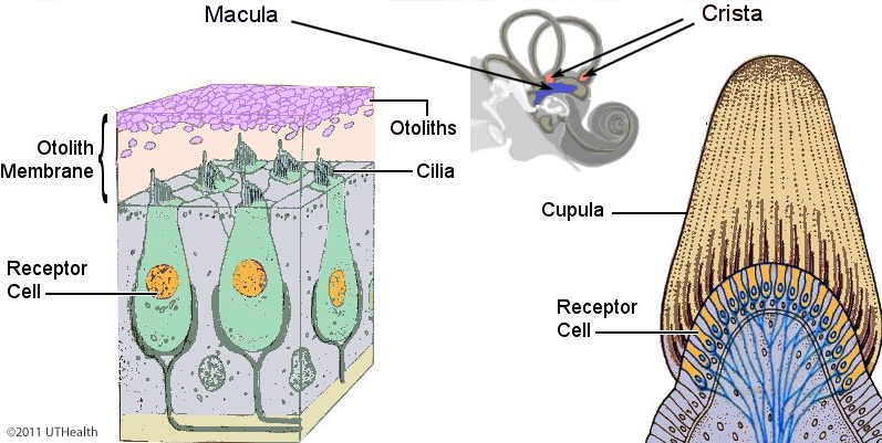
The crista or ampullary crest is a transverse ridge of connective tissue, blood vessels and nerve fibers that is located in each ampulla of the three semicircular ducts. The surface of the crista consists of a thin layer of receptor cells (vestibular hair cells) and supporting cells. A clear gel-like, dome-shaped mass, the cupula, normally rests on the crista surface and extends up to the roof of the ampulla. Although you cannot see the receptor cells in any detail in this figure, you should be aware of their general structural features. Receptors of the vestibular organ are hair-bearing specialized epithelial cells similar to the auditory receptor cells. These receptor cells contain synaptic vesicles and generate receptor potentials when the hairs are deflected by mechanical (vestibular) stimuli. About 40 to 80 stereocilia and a single kinocilium project from the upper surface of these cells. The stereocilia are arranged in rows which show a progressive increase in length. The single kinocilium is located next to the row containing the longest stereocilia. The stereocilia and the kinocilium of the crista hair cells are embedded in the cupula. Deflection of the cupula by endolymph flow and the resulting bending of the stereocilia and kinocilium are believed to stimulate these hair cells.
The macula of the saccule consists of a thickened layer of connective tissue, blood vessels and nerve fibers that is covered by a thin layer of receptor and supporting cells. In microscopic images the receptor surface of the macula appears as a dark band, and tufts of cilia can be seen extending from the receptor surface (the dark band) into the otolith membrane (the light band). The receptor cells in the macula are identical to those in the crista; each cell bears numerous stereocilia and a single kinocilium and contains synaptic vesicles. The macula differs from the crista by being covered by a flattened layer of gelatinous matter, the otolith membrane, (the light band above the receptor surface) which is covered, in turn, by small crystals of calcium carbonate called the otoliths (the dark clumps resting on the membrane). The cilia of the macular receptor cells are embedded in the otolith membrane. It is believed that the effective stimulus to the macular hair cells is the bending of the hairs that occurs when the otoliths are shifted during linear acceleration or deceleration and during head tilt. The macula of the utricle is similar to but not identical to the macula in the saccule.