Lab 7 (ƒ6) - Auditory, Vestibular, Gustatory and Olfaction Systems
Midbrain and Diencephalon
View and identify the structures in the illustration.
Figure 1, Layer A is a section through the midbrain at the level of the inferior colliculus. Identify the nucleus of the inferior colliculus. Notice the periaqueductal gray surrounds the cerebral aqueduct; the inferior colliculus which forms the tectum, and the decussation of the superior cerebellar peduncles. The corticofugal fibers in thecrus cerebri form the base of the midbrain. The substantia nigra marks the floor of the midbrain tegmentum. The medial lemniscus, ventral trigeminal lemniscus, and spinothalamic tract are starting to sweep posterolaterally on their course to the thalamus. Find the mesencephalic trigeminal tract and nucleus. The fibers of the lateral lemniscus are terminating within the inferior colliculus. The axons of the inferior colliculus (3°, 4° and 5° afferents) are collecting along the lateral edge of the inferior colliculus to form the brachium of the inferior colliculus.
Figure 1, Layer B shows the somatosensory tracts (medial lemniscus, ventral trigeminal lemniscus, spinothalamic tract) displaced posterolaterally by the red nucleus. The superior colliculus now forms the midbrain tectum and is posterior to the cerebral aqueduct. The periaqueductal gray continues to surround the cerebral aqueduct. Thesubstantia nigra forms the anterior border of the midbrain tegmentum and is posterior to the crus cerebri. Observe the fascicles of the oculomotor (III) nerve leaving the brain stem within the interpeduncular fossa.
The brachium of the inferior colliculus (3° to 5° auditory afferents) is located lateral to the somatosensory tracts and is entering the medial geniculate body (MGB) of the thalamus. The axons of the MGB collect along its lateral margin to form the auditory radiations or geniculotemporal tract, which travel in the sublenticular segment of the internal capsule. These axons terminate in the superior bank of the superior temporal gyrus.
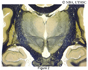 Figure 2, is a coronal section through the diencephalon and midbrain. Notice that the internal capsule posterior limb is located at the lateral border of the thalamus and that the third ventricle is located between the two thalami. The small dark fiber band on the medial surface of the thalamus is the stria medullaris thalami. Recall that the sublenticular limb of the internal capsule is located inferior to the lenticular nucleus. Notice that in this section you can see segments of the diencephalon (i.e., the thalamus) and the midbrain (i.e., the red nucleus, substantia nigra and crus cerebri).
The axons of the MGB travel rostrally and laterally to form the auditory radiations or geniculotemporal tract within the sublenticular segment of the internal capsule. These fibers terminate in the superior bank or transverse gyrus of the superior temporal lobe. Recall that in the gross specimen, the expanse of the transverse gyrus of the superior temporal lobe could only be appreciated by separating the "lips" of the lateral fissure. The primary auditory cortical receiving area forms the lower "lip" of the lateral fissure. Electrical stimulation of cortical areas in and near the primary auditory area in unanesthetized subjects produces the sensations of sounds described as that of a bell or a whistle. Most of these "sounds" are referred to the contralateral ear. Because the auditory system is bilaterally represented at and above the level of the superior olivary complex, damage of the auditory cortex results in only a partial hearing deficit. The deficits are bilateral, with the greatest loss contralateral to the lesion. Consequently, unilateral damage to the auditory cortex is difficult to recognize.