Lab 7 (ƒ6) - Auditory, Vestibular, Gustatory and Olfaction Systems
The Auditory System - Introduction
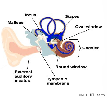
The auditory system is an exteroceptive system involved in the perception of sound. The receptor organ includes the outer, middle and inner ears. The outer ear consists of the pinna and external auditory canal or meatus. The middle ear is an air-filled bony chamber which is partitioned from the outer ear by the tympanic membrane or ear drum. The middle ear ossicles (a chain of three small bones in the middle ear cavity) conduct sound energy from the tympanic membrane to the inner ear.
 The auditory portion of the inner ear is a fluid-filled bony chamber, the cochlea, which contains a membranous, fluid-filled cochlear duct. The receptor cells are located in the organ of Corti within the cochlear duct. The organ of Corti consists of supporting and sensory receptor cells. A prominent feature of the organ of Corti is the space called the tunnel of Corti, which separates two groups of hair receptor cells. The hair receptor cells are arranged in two groups, a row of inner hair cells near the osseous spiral lamina and a band of outer hair cells organized in three rows.
The modiolus is the bony core of the cochlea around which the cochlear canal and cochlear duct spiral. Located within the modiolus are the bipolar cell bodies of the 1° afferents which constitute the spiral ganglion. The auditory nerve, the central processes of the 1° afferents, collect within and pass through the modiolus. The cochlear canal is a bony channel that makes two and one-half turns (in humans) around the modiolus. Notice that the cochlear duct is suspended within the cochlear canal and encloses a chamber, the scala media. This chamber contains the fluid endolymph. In other words, the walls of the chamber are formed by the cochlear duct and the enclosed space is the scala media. Notice that the cochlear duct divides the cochlear canal into three chambers (except at the helicotrema.) The largest chamber is the scala tympani, the middle chamber is the scala media and the third chamber is the scala vestibuli.
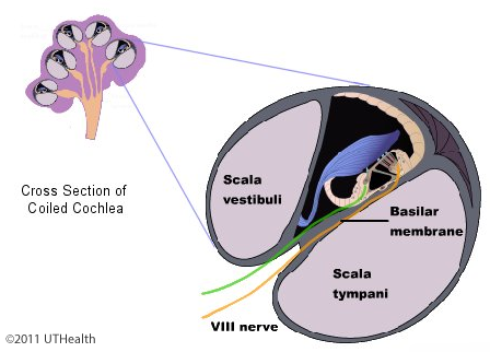 The cochlear duct has a triangular shape. Its three walls are formed by the basilar membrane, the stria vascularis, and the vestibular (Reissner's) membrane. The bony cochlear canal is divided in half by the osseous and membranous spiral lamina. The osseous spiral lamina is a bony ridge extending from the modiolus into the cochlear canal. The membranous spiral lamina or basilar membrane is a fairly thick membrane which extends from the osseous spiral lamina to the outer cochlear wall.
The vestibular membrane is a thin sheet of tissue extending obliquely from the spiral limbus to the outer cochlear wall. The spiral limbus is formed by a thickening of the soft tissue overlying the osseous spiral lamina.
It is easy to identify the cochlear chambers if you remember that the vestibular membrane (the thinnest wall of the cochlear duct) separates the scala vestibulifrom the scala media. The stria vascularis is a highly vascularized, pigmented cell layer lining the outer cochlear wall between the points of attachments of the basilar and vestibular membranes. The stria vascularis is believed to be important in maintaining the unique ionic content of the endolymph.
Extending peripherally from the spiral limbus, over the organ of Corti, is the tectorial membrane. This membrane has a gelatinous surface facing the receptor hair cells. It is believed that the hairs of the receptors are embedded within this surface. Although you cannot clearly visualize the receptor cells you should be aware of their general structure. The receptor cells are modified epithelial cells that contain synaptic vesicles and that generate receptor potentials to acoustic stimuli. The receptor cells are oblong (outer hair cells) of globular (inner hair cells) in shape and at their apical end, bear cilia or hairs. The hairs line up to form the shape of a W or U. These hairs are embedded in a cuticular layer at the upper end of the hair receptor cell. I humans, the air cells are arranged in three rows of outer hair cells and one row of inner hair cells. The terminals of the peripheral processes of the first - order afferents end near the base and lower third of the receptor cell. Efferent terminals of brainstem neurons are also found synapsing upon the outer hair cells. Efferents do not appear to synapse directly upon inner hair cells, but end on the afferent fibers innervating the inner hair cells. Both types of efferent terminals are part of neural feedback circuits that permit brainstem structure to control the inflow of auditory information. These efferents are collectively called the olivocochlear bundle. Within a canal, the bony modiolus of the cochlea, the cell bodies of the auditory primary afferents form a spiral -shaped ganglion called the spiral ganglion. These neurons can be seen in the base of the osseous spiral lamina.