Lab 8 (ƒ7) - Visual System
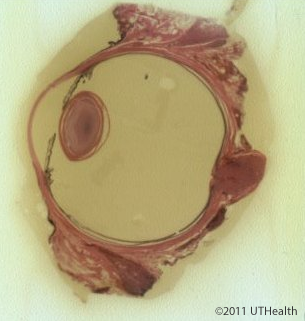
Microscopic Anatomy of the Eye
 The regulation of pupil size and lens curvature involves the autonomic nervous system. Stimulation of the cranial parasympathetic system results in the constriction of the pupil and increased lens curvature, while stimulation of the spinal sympathetic system results in the dilation of the pupil.
The cranial or parasympathetic motor pathway involved in pupil constriction and in lens accommodation includes the Edinger-Westphal nucleus of the oculomotor nuclear complex (the preganglionic parasympathetic neuron), the oculomotor nerve (in which the preganglionic parasympathetic fibers travel), and the ciliary ganglion (the postganglionic parasympathetic neuron). The postganglionic axons travel in the short ciliary nerve to the sphincter muscle of the iris and to the ciliary muscle of the ciliary body. (Recall that the ciliary muscles control lens position and curvature.) The spinal or sympathetic motor pathway involved in pupil dilation includes lateral horn cells of the upper thoracic segments (the preganglionic sympathetic neurons), the anterior root (in which the preganglionic sympathetic fibers leave the spinal cord), and the superior cervical ganglion (the postganglionic sympathetic neurons). The postganglionic sympathetic fibers terminate on the dilator muscle of the iris.