6.1 Amygdala - General Considerations
Amygdala is the integrative center for emotions, emotional behavior, and motivation. If the brain is turned upside down the end of the structure continuous with the hippocampus is called the uncus. If you peel away uncus you will expose the amygdala which abuts the anterior of the hippocampus. Just like with the hippocampus, major pathways communicate bidirectionally and contain both efferent and afferent fibers.
Figure 6.1 |
6.2 Inputs to the Amygdala
Figure 6.2 |
As was the case with the hippocampus, fibers carrying inputs to the amygdala are in virtually all cases combined with fibers carrying outputs from the amygdala.
The amygdala receives inputs from all senses as well as visceral inputs. Since the amygdala is very important in emotional learning it is not surprising that visceral inputs are a major input source. Visceral inputs come from the hypothalamus, septal area, orbital cortex, and parabrachial nucleus. Olfactory sensory information comes from the olfactory bulb. Auditory, visual and somatosensory information comes from the temporal and anterior cingulate cortices.
Figure 6.3 |
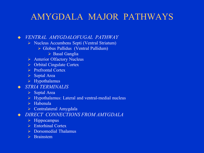
6.3 Major Output Pathways of the Amygdala
- Ventral amygdalofugal pathway
- Stria terminalis
- Directly to the hippocampus
- Directly to the entorhinal cortex
- Directly to the dorsomedial nucleus of the thalamus
6.4 Ventral Amygdalofugal Pathway
Ventral Amygdalofugal Pathway. The term "fugal" comes from the word fuge—to drive away—as in fugitive. This pathway continues to the anterior olfactory nucleus, anterior perforated substance, piriform cortex, orbitofrontal cortex, anterior cingulate cortex, and ventral striatum. The ventral striatum includes part of the caudate, putamen, and the nucleus accumbens septi (nucleus that reclines on the septum). Projections from the ventral striatum are links in a basal ganglia circuit that are important in stimulus-response associative learning. The ventral amygdalofugal pathway also connects to the hypothalamus and septal nucleus, but the amygdala's major connection to the hypothalamus and septal nucleus is through the stria terminalis.
The ventral amygdalofugal pathway is important because it is a link whereby motivation and drives, through the limbic system, can influence responses. It is also a link whereby responses are learned. In this case this is the link whereby associative learning takes place. That is where responses are associated with appetitive and aversive consequences that is rewards and punishers.
Three simplifications:
- The stria terminalis is similar in form, function, and location as the fornix for the hippocampal pathway. Thus by way of analogy one can say that the stria terminalis is to the amygdala as the fornix is to the hippocampus. Stria is a Latin word that means line, groove, or band. Related to the word "striated".
- The stria terminalis connects only to subcortical structures. (Connection to cortical structures is through the ventral amygdalofugal pathway.)
- The stria terminalis overlaps with the ventral amygdalofugal pathway in that it also connects to the septal nuclei and hypothalamus and thus forms a loop.
More on similarities to the fornix:
Like the fornix, the stria terminalis has precommissural and postcommissural branches in relation to the anterior commissure. The precommissural branch goes to the septal area. This is exactly what the fornix does. The postcommissural branch goes to the hypothalamus. This is exactly what the fornix does. Whereas the postcommissural branch of the fornix projects to mammillary bodies of the hypothalamus, the postcommissural branch of the stria terminalis projects to the lateral nucleus and ventral-medial nucleus of the hypothalamus.
As with the fornix, some fibers enter anterior commissure cross to the contralateral side. Just as in the case of the two hippocampi communicating with each other through the anterior commissure, the two amygdala communicate with each other through the anterior commissure.
The stria terminalis also projects to the habenula, which is part of the epithalamus.
The central nucleus of the amygdala produces autonomic components of emotion (e.g., changes in heart rate, blood pressure, and respiration) primarily through output pathways to the lateral hypothalamus and brain stem.
The central nucleus of the amygdala also produces conscious perception of emotion primarily through the ventral amygdalofugal output pathway to the anterior cingulate cortex, orbitofrontal cortex, and prefrontal cortex.
6.5 More on Function of the Amygdala
Stimulation of the amygdala causes intense emotion, such as aggression or fear.
Irritative lesions of temporal lobe epilepsy have the effect of stimulating the amygdala. In its extreme form irritative lesions of temporal lobe epilepsy can cause a panic attack. Panic attacks are brief spontaneously recurrent episodes of terror that generate a sense of impending disaster without a clearly identifiable cause. PET scans have shown an increase in blood flow to the parahippocampal gyri, beginning with the right parahippocampal gyrus. Similar but attenuated blood flow increases occurs during anxiety attacks.
Destructive lesions such as ablation of the amygdala cause an effect opposite to the irritative lesions of temporal lobe epilepsy. Destructive lesions of the amygdala cause tameness in animals, and a placid calmness in humans characterized as a flatness of affect. Lesions of the amygdala can occur as a result of Urbach-Wiethe disease where calcium is deposited in the amygdala. If this disease occurs early in life then these patients with bilateral amygdala lesions cannot discriminate emotion in facial expressions, but their ability to identify faces remains. The anatomical area for face recognition and memory is in the multimodal association area of the inferotemporal cortex. This is a good example of how emotion in one area (amygdala) is linked with perception in another area (inferotemporal cortex) to create an intense emotionally charged memory.
Figure 6.4 |
Flatness of affect is one of the symptoms of the previously mentioned Kluver-Bucy syndrome where the entire temporal lobes of monkeys were removed. Actually,just lesions of the amygdala were shown to be primarily responsible for flatness of affect. This work eventually led to the psychosurgical technique of prefrontal lobotomies. Remember the movie with Jack Nicholson, “One Flew Over the Cuckoo’s Nest.” The prefrontal cortex inputs into the amygdala. By severing this input a flatness of affect is produced which was thought to be desirable in schizophrenic patients who were aggressively violent or emotionally agitated.
The amygdala combines many different sensory inputs. Like the hippocampus it combines external and internal stimuli. Every sensory modality has input. These are integrated with somatosensory and visceral inputs—this is where you get your “gut reaction”. The link between prefrontal cortex, septal area, hypothalamus, and amygdala likely gives us our gut feelings, those subjective feelings, about what is good and what is bad.
It is also where memory and emotions are combined. When the reward is particularly sweet, that behavior and association may last a lifetime. Likewise, the trauma and humiliation of punishment may be remembered for a long time too.
6.6 Fear Conditioning: An Example of the Role of the Amygdala in Learning
Another example of emotion being linked to some perceptual experience is fear conditioning. In this example the sensory experience is auditory rather than visual as in the emotion of faces. Much of what we know about the amygdala and its role in emotional learning and memory comes from fear conditioning, mostly but not exclusively conducted with animals. This is an example of classical conditioning or Pavlovian conditioning. In the classic experiments conducted by Pavlov just after the turn of the century, a neutral stimulus—a bell—was sounded and after a brief interval food powder—the unconditioned stimulus—was placed in the dog’s mouth. After a few such pairings the dog would salivate to the sound of the bell. The crucial aspect of classical conditioning is that it is a pairing between two stimuli. No response is required to get the reward. In fear conditioning, an organism hears a noise or sees a visual stimulus. A few seconds, later it receives a mild shock. The reactions involve freezing, elevated blood pressure and heart rate, and it gets twitchy—startles easily.
Figure 6.5
|
|
|
Figure 6.6 (top) and 6.7 (bottom) |
Pathways from the thalamus to the amygdala are particularly important in emotional learning. Output pathways from the central nucleus of the amygdala make extensive connections with the brain stem for emotional responses and extensive connections with cortical areas through the nucleus basalis. Cholinergic projections from the nucleus basalis to the cortex are thought to arouse the cortex.
The following diagram provides additional information on outputs controlled by the amygdala during fear conditioning.
Figure 6.8 |
Some pathways of fear conditioning have been discovered and this is a hot research topic in neuroscience. If the auditory cortex pathway is lesioned, for example, basic fear conditioning is unaltered, but discrimination is altered. In the discrimination procedure one sound is paired with shock and another sound is not paired with shock. The animals had to rely solely on the thalamus and amygdala for learning and they could not learn the discrimination; apparently the two stimuli were indistinguishable.
So, the cortex is not needed for simple fear conditioning; instead it allows us to recognize an object by sight or sound— to interpret the environment.
Thus, pathways from the sensory thalamus provide only a crude perception of the world, but because they involve only one neural link they are fast pathways. Why might FAST be important? We need a quick reaction to potential danger. The thalamus—amygdala pathway provides us with this and may also prepare the amygdala to receive more highly processed information from the cortex.
On the other hand, pathways from the cortex offer detailed and accurate representations of the environment. Because these pathways have multiple neural links they are slow by comparison.
If for example we see a slender curled shape behind a tree its much better to jump back and later recognize its a garden hose than to fail to quickly jump back if it were a snake. There is plenty of time later to reflect that it was foolish to be startled in our own secure garden where there are no snakes.
Figure 6.9 |
Figure 6.10 |
Cortical vs. subcortical pathways of fear conditioning. |
|
Fear producing visual stimuli is quickly processed by the thalamus and this information is passed to the amygdala (red) producing a quick response (green) to danger. The thalamus also passes the information to the cortex so that more careful (and slower) judgments can be made about the real potential danger.
The amygdala is involved in pleasureful emotional learning as well as fearful emotional learning. Consider instrumental learning. Unlike classical conditioning where two stimuli are paired, in instrumental conditioning responses are followed by reward and stimulus-response associations are learned. There are thus three events: a stimulus, a response, and a reward. It has become clear that all three pairwise combinations are learned in instrumental conditioning. Where the amygdala comes in is that lesions of the basolateral nuclei of the amygdala disrupt the association the stimulus and rewarding attributes of the food.
This amygdala memory system serves as an example of memory systems generally. The establishment of memories is a function of the entire network, not any single component. The amygdala is involved in a kind of primitive emotional memory, one that is likely preserved by evolution. According to the diagram of memory systems (e.g., Nolte, p.577), declarative memory is mediated by the hippocampus and the cortex. But like the cortex, lesions of the hippocampus have little effect on fear conditioning except in discriminating environmental stimuli.
A study of patients with damage to the amygdala, hippocampus, or both clearly demonstrates the distinctive roles of these two structures in memory. These patients were shown slides of green, blue, yellow, or red colors. After some colors, a loud and frightening horn blast was sounded. Autonomic responses were recorded (via GSR recordings) to determine learning. Amygdala patients did not become conditioned to colors followed by the loud horn. But when asked how many colors were presented and which were followed by the horn, their recall was correct. That is, they had explicit memory about the events. On the other hand, hippocampal patients showed learning and conditioning to the colors followed by the horn, but could not recall which they were. That is, they had implicit memory about the events. Patients with both types of lesions showed no conditioning and had no explicit memory about which colors were followed by the horn. The chapter on Learning and Memory will explain more about explicit memory and the hippocampus.