The analysis of the anatomical and physical bases of learning and memory is one of the great successes of modern neuroscience. Thirty years ago little was known about how memory works, but now we know a great deal. This Chapter will discuss four issues that are central to learning and memory. First, what are the different types of memory? Second, where in the brain is memory located? One possibility is that human memory is similar to the memory chip in a personal computer (PC), which stores all the memory in one location. A second possibility is that our memories are distributed and stored in different regions of the brain. Third, how does memory work? What types of changes occur in the nervous system when a memory is formed and stored, are there particular genes and proteins that are involved in memory, and how can a memory last for a lifetime? Fourth, is the issue of importance to many people, especially as we age: How can memory be maintained and improved, and how can it be fixed when it is broken?
7.1 Types of Memory
Psychologists and neuroscientists have divided memory systems into two broad categories, declarative and nondeclarative (Figure 7.1). The declarative memory system is the system of memory that is perhaps the most familiar. It is the memory system that has a conscious component and it includes the memories of facts and events. A fact like 'Paris is the capital of France', or an event like a prior vacation to Paris. Nondeclarative memory, also called implicit memory, includes the types of memory systems that do not have a conscious component but are nevertheless extremely important. They include the memories for skills and habits (e.g., riding a bicycle, driving a car, playing golf or tennis or a piano), a phenomenon called priming, simple forms of associative learning [e.g., classical conditioning (Pavlovian conditioning)], and finally simple forms of nonassociative learning such as habituation and sensitization. Sensitization will be discussed in detail later in the Chapter. Declarative memory is "knowing what" and nondeclarative memory is "knowing how".
Figure 7.1 |
7.2 Testing Memory
Figure 7.2 |
Figure 7.3 |
Everyone is interested in knowing how well they remember so let us take a simple memory test. The test (Figure 7.2) will present a list of 15 words, then there will be a pause and you will be asked whether you remember some of those words. Sorry, you have to put your pen down for this test and do not read further in the Chapter until you complete the test.
This memory test called the DRM test after its creators James Deese, Henry Roediger and Kathleen McDermott. It was not meant to be a trick, but to illustrate a very interesting and important feature about memory. We like to think that memory is similar to taking a photograph and placing that photograph into a filing cabinet drawer to be withdrawn later (recalled) as the “memory” exactly the way it was placed there originally (stored). But memory is more like taking a picture and tearing it up into small pieces and putting the pieces in different drawers. The memory is then recalled by reconstructing the memory from the individual fragments of the memory. The reason so many individuals incorrectly believe that “sweet” was on the list is because there were so many other words on the list that had a sweet connotation. “Failing” this test is actually not a bad outcome. Individuals with Alzheimer’s disease generally do not say that “sweet” was on the list. They cannot make the normal associations involved in the recall of a memory.
The word list gives insights into memory processing and retrieval, but it is not a really good test of “raw” memory ability because it can be affected by distortions and biases. To avoid these problems, psychologists have developed other memory tests. One is the object recognition test (Figure 7.3) to test declarative memory. This test is also good because, as we will see later, it can even be used on animals. The test involves presenting a subject with two different objects and they are asked to remember those objects. There is a pause and then two objects are shown again, one of which is new and the other having been shown previously. Subjects are asked to identify the novel object, and to do so, they need to remember which one was shown previously. A somewhat related test is the object location test in which subjects are asked to remember the location of an object on a two-dimensional surface.
Examples of nondeclarative memory, such as associative learning, can be tested by pairing one stimulus with another and later testing whether a subject has learned to make the association between the two stimuli. The classical example is the paradigm developed by the Russian physiologist Ivan Pavlov, which is now called classical or Pavlovian conditioning. In classical conditioning (Figure 7.4), a novel or weak stimulus (conditioned stimulus, CS) like a sound is paired with a stimulus like food that generally elicits a reflexive response (unconditioned response, UR; unconditioned stimulus, US) such as salivation. After sufficient training with contingent CS-US presentations (which may be a single trial), the CS is capable of eliciting a response (conditioned response, CR), which often resembles the UR (or some aspect of it).
Figure 7.4 |
7.3 Localization of Memory
Now let us turn to this issue about where is memory located. There are three basic approaches.
- Imaging. Modern imaging techniques like fMRI (functional magnetic resonance imaging) or PET (positron emission tomography) allows one to “see” areas of the brain that are active during specific brain tasks. If a subject is placed in an fMRI scanner and given a memory test, one can determine what areas of the brain are active, and that activity presumably is related to where in the brain the memory is processed and/or stored.
Figure 7.5
PET brain scan during an object location test. (from A. M. Owen, et al., J. Cog. Neurosci. 8:6, 588-602, 1996.)
Figure 7.5 illustrates an example of a PET scan of an individual who is performing an object location test. The color code is such that the brighter, redder regions indicate increased brain activity. The most active region is the hippocampus. In discussions of memory, the hippocampus is mentioned repeatedly because it is a major part of the brain involved in declarative memory function. This illustration clearly indicates that the hippocampus is involved in object location memory. But as we will see soon, it is not where all memories are stored.
- Brain lesions. In this experimental procedure, small parts of the brains of mice or rats are surgically removed or chemically inactivated and the animals are systematically examined to determine whether the lesion affected any memory system.
- Brain disease and injury. Here scientists take advantage of individuals who have had unfortunate brain injuries, for example, through stroke or through a brain tumor in a specific area of the brain. If one finds a memory deficit in the patient, it is likely that the region of the brain that was injured is involved in that memory.
A classic study on localization of memory was the result of surgery performed on Henry Molaison, a patient who was only known to the scientific community as “H.M.” until his death in 2008. H. M. is famous in neuroscience literature because his brain provided major insights into the localization of memory function. In the 1950’s, H.M. was diagnosed with intractable epilepsy, and while there are pharmacologic treatments, in some cases the only treatment is to remove the portion of the brain that is causing the seizures. Consequently, H.M.'s hippocampus was removed bilaterally. Figure 7.6 (right) is an MRI of a normal individual showing the hippocampal region, whereas Figure 7.6 (left) shows a MRI of patient H.M. after the removal of the hippocampus.
Figure 7.6
Bran scans of H.M. (left), and a normal individual (right). (Copyright © 1997 by Suzanne Corkin, used with permission of The Wylie Agency LLC.)
Before the operation, H.M. had a fine memory, but after the operation, H.M. had a very severe memory deficit. Specifically, after the operation H.M.'s ability to form any new memories for facts and events was severely impaired; he had great difficulty learning any new vocabulary words; he could not remember what happened the day before. So if H.M. had an interview the day following a previous interview, he would have little or no memory about the interview or events during it. This study clearly indicated that the hippocampus was critical for memory formation. But whereas H.M. had great difficulty forming new memories for facts and events, he still had all of his old memories for facts and events. Specifically, he had all his childhood memories, and all of his memories prior to the operation. This type of memory deficit is called anterograde amnesia. (In contrast, retrograde amnesia refers to loss of old memories.) The studies on H.M. clearly indicated that whereas the hippocampus is critical for the formation of new memories, it is not where the old memories are stored. It is now known that those old memories are stored in other parts of the brain, such as in the frontal cortex. The process by which an initially labile memory is transformed into a more enduring form is called consolidation. This process involves the memory being stored in a different part of the brain than the initial site of its encoding.
H.M. was also interesting in that while his ability to form new memories for facts and events was severely impaired, he could form new memories for skills and habits. While he could form new memories for skills and habits, he did not know that he had the skills! He had no awareness of the memory; he couldn’t declare that he had it. This finding clearly indicated that the memory for skills and habits are not formed in the hippocampus. Collectively, we learned from these studies on H.M. and other patients that memory is distributed throughout the nervous system, and different brain regions are involved in mediating different types of memory.
Figure 7.7 summarizes many decades of research on the anatomical locus of memory systems. The medial temporal lobe and structures like the hippocampus are involved with memories for facts and events; the striatum is involved with memories for skills and habits; the neocortex is involved with priming; the amygdala is involved with emotional memories; and the cerebellum with simple forms of associative learning. Lower brain regions and the spinal cord contain even simpler forms of learning. In summary, memory is not stored in a single place in the brain. It is distributed in different parts of the brain.
Figure 7.7 |
7.4 Mechanisms of Memory
Model systems to study memory mechanisms
Much of what has been learned about the neural and molecular mechanisms of learning and memory have come from the use of so called “model systems” that are amenable to cellular analyses. One of those model systems is illustrated in Figure 7.8A. Aplysia californica is found in the tidal pools along the coast of Southern California. It is about six inches long and weighs about 150 grams. At first glance it is an unpromising looking creature, but neuroscientists have exploited the technical advantages of this animal to gain fundamental insights into the molecular mechanisms of memory. Indeed, the pioneering discoveries of Eric Kandel using this animal were recognized by his receipt of the Nobel Prize in Physiology or Medicine in 2000. Aplysia have three technical advantages.
First, it exhibits simple forms of nondeclarative (implicit) learning like classical (Pavlovian) conditioning, operant conditioning and sensitization.
Second, Aplysia have a very simple nervous system. Compared to the 100’s of billions of nerve cells in the human brain, the entire nervous system of this animal only has about 10,000 cells. Those cells are distributed in different ganglia like the one illustrated in Figure 7.8B. Each ganglia like this one has only about 2,000 cells, yet it is capable of mediating or controlling a number of different behaviors. This means that any one behavior can be controlled by 100 neurons or even less. One has the potential of working out the complete neural circuit underlying a behavior, and then, after training the animal, the neural circuit can be examined to identify what has changed in the circuit that underlies the memory.
Third, the ganglia contain neurons that are very large. Figure 7.8B shows a ganglion under a dissecting microscope. It is about 2mm in diameter. The spherical structures throughout the ganglia are the cell bodies of individual neurons. Each neuron is identifiable and has a unique localization and function. A related advantage is that individual neurons can be removed and placed in culture medium where they can survive for many days. Indeed, multiple neurons can be removed from the ganglia and they reestablish their normal synaptic connections, thereby providing a very powerful experimental system to study the physiology of nerve cells and the properties of the connections between them. Figure 7.8C shows an example of a sensory neuron (small cell to the right) and a motor neuron (large cell to the left) in culture. In the micrograph it is possible to see the shadow of a microelectrode that has impaled the sensory neuron, and the shadow of a microelectrode that has impaled a motor neuron for performing intracellular recordings.
Sensitization, a simple form of nondeclarative learning amenable to detailed cellular analyses
Figure 7.9 |
| A. | B. | C. |
Figure 7.10 |
||
Figures 7.9 and 7.10 illustrate a simple behavior exhibited by the animal and a simple form of learning called sensitization. The animal is tested by stimulating its tail with a weak electric shock (7.9) or a weak mechanical tap (7.10). These stimuli elicit defensive reflex withdrawals of the body, which includes the tail and nearby sites such as the gill and a fleshy spout called the siphon. In response to test stimuli delivered every five minutes, the withdrawals are fairly reliable. They are about the same duration each time (Figures 7.9B, C, 7.10A). But if a strong noxious stimulus (e.g., an electric shock) is delivered to another part of the animal such as its body wall, subsequent test stimuli to the tail give enhanced responses (Figure 7.9B and 7.10B). This is an example of a simple form of learning called sensitization. It is defined as the enhancement of the response to a test stimulus as a result of delivering a strong generally noxious stimulus to the animal. In a sense, the animal is learning that it is in a “fearful” environment. Sensitization is a ubiquitous form of learning that is exhibited by all animals including humans.
Neural circuit and mechanisms of sensitization
- Neural circuit. We can take advantage of the large nerve cells of Aplysia, and the ability to make intracellular recordings from them, to work out the underlying neural circuit. Figure 7.11 illustrates a simplified view of the key components of the underlying neural circuit. Stimulation of the skin activates sensory neurons (SN) (only one of which is illustrated here) which make glutamatergic excitatory synaptic connections (triangles) with motor neurons (MN). If the summated synaptic input to the motor neurons is sufficiently large, the motor neurons will be activated and action potentials will propagate out of the ganglion to cause an eventual contraction of the muscle. So stimulation of the skin excites sensory neurons, the sensory neurons activate motor neurons, and motor neurons contract the muscles. Also, it should be evident that the greater the activation of the motor neurons, the greater will be the subsequent reflex response. This reflex in Aplysia is similar to the knee jerk or stretch reflex mediated by similar circuitry in the vertebrate spinal cord.
Figure 7.11
Neural circuit for the defensive withdrawal reflex.
- Mechanisms of sensitization. Sensitizing stimuli lead to the release of the neurotransmitter serotonin (5-HT) (represented by cell labeled IN and colored in purple on Figure 7.11). 5-HT modulates the strength of the connection between the sensory neuron and the motor neuron. An action potential in the sensory neuron before the learning produces a small excitatory postsynaptic potential (EPSP) in the motor neuron (Figure 7.12A). But, after delivering the sensitizing stimulus, an action potential in the sensory neuron leads to a larger synaptic potential in the motor neuron (Figure 7.12C). A larger synaptic potential in the motor neuron increases the probability that the motor neuron will be activated to a greater extent and produce a larger contraction of the muscle (i.e., sensitization).
One principle about learning and memory derived from studies of this simple animal, and this principle holds true in our brains as well, is that learning involves changes in the strength of synaptic connections between neurons. Learning is not due to a reorganization of the nervous system or the growth of new neurons. What has changed is that the strength of a previously existing connection is modified.
Now we can take this analysis one step further and ask what are the biochemical mechanisms that underlie learning and memory. We will divide the discussion into two temporal domains of memory; short-term memory and long-term memory. We have already discussed different types of memory such as declarative and nondeclarative memory. There are also different temporal domains of memory. Short-term memories are like the memory for a telephone number that last several minutes, and long-term memory are memories that last days, weeks or a lifetime.
- Mechanisms of short-term sensitization. The mechanisms for the short-term memory for sensitization are illustrated in Figure 7.12B. The sensitizing stimulus leads to release of the neurotransmitter 5-HT. 5-HT binds the two types of receptors on the sensory neuron; one is coupled to the DAG/PKC system, and the other is coupled to the cyclic AMP/PKA system. These are the same general cascades that you learned in biochemistry. Learning mechanisms have evolved to co-opt some of the biochemical machinery that are already present in all cells used them specifically for a memory mechanism in nerve cells. The protein kinases exert two types of actions. First, they regulate the properties of different membrane channels (the small gates on the illustration (Figure 7.12) represent membrane channels that underlie the initiation and the repolarization of the action potential). Consequently after a sensitizing stimulus, the amount of calcium that enters the synaptic terminal during an action potential and causes the release of transmitter will be enhanced. In addition, the modulation of the membrane channels leads to an increase in the excitability of the sensory neuron and as a result a greater number of action potentials will be elicited by a test stimulus to the skin. Second, the kinases regulate other cellular processes involved in transmitter release, such as the size of the pool of synaptic vesicles available for release in response to the influx of Ca2+ with each action potential. Finally, 5-HT leads to changes in the properties of the postsynaptic motor neuron. Specifically, 5-HT leads to an increase in the number of glutamate receptors. The consequences of these processes can be seen by comparing the strength of the synaptic connection produced by a single action potential before (Figure 7.12A) and after (Figure 7.12C) sensitization. The specific details of all the currents and processes are not critical. However, it is important to know the general principles. One principle is that learning involves the engagement of second messenger systems. Here both the protein kinase C (PKC) and the protein kinase A (PKA) systems are involved. This is a fairly general principle. In every example of learning that has ever been examined, whether vertebrate or invertebrate, second messenger systems are engaged. A second principle is that memory involves the modulation of neuronal membrane channels. These can include channels that directly regulate transmitter release (i.e., Ca2+ channels in the presynaptic neuron), channels that regulate neuronal excitability, and channels that mediate synaptic responses in the postsynaptic neuron. A third principle is that cyclic AMP is one of the critical second messengers that is involved in memory. Given this information, you can begin to think about how memory could be improved based on your knowledge of the underlying biochemistry.
We have discussed a mechanism for a short-term memory. It is "short-term" because the memory is transient and that is so because the underlying biochemical changes are transient. The duration of the memory is dependent on how long the various substrate proteins (e.g., membrane channels) are phosphorylated. PKA will only be activated for a short time after a brief stimulus because cyclic AMP will be degraded and PKA levels will decrease. Protein phosphatases will remove the phosphate groups on the substrate proteins that are “storing” the memory.
Figure 7.13
Structural changes in sensory neurons associated with long-term sensitization. (Modified from M. Wainwright et al., J. Neurosci. 22:4132-4141, 2002.)
- Mechanisms of long-term sensitization. There are two major differences between short-term and long-term memories. Long-term memories involve changes in protein synthesis and gene regulation, whereas short-term memories do not. And, long-term memories in many cases involve structural modifications. Figure 7.13 illustrates examples of the processes of two sensory neurons that have been filled with a dye, one from an untrained animal and one from a trained animal. Shown are the thick axonal process of the neuron and many fine branches. Along the branches are seen small dot-like swellings or varicosities. These varicosities are the presynaptic terminals of the sensory neurons that make contact with other neurons like the motor neurons. (The motor neurons cannot be seen because only the sensory neurons were filled with the dye.) Part B of Figure 7.13 shows an example of a sensory neuron that has been injected with a dye in an untrained animal, and Part A shows one that has been filled with a dye 24 h after sensitization training. There is a major difference between these two neurons. The neuron from the trained animal has a greater number of branches and a greater number of synaptic varicosities than the neuron from the untrained animal. Therefore, long-term memory involves changes in the structure of neurons including growth of new processes and synapses. So, to the extent that you remember anything about this material on memory tomorrow, or next week, or next year, it will be because structural changes in synapses are beginning in your brains!
Figure 7.14
Genes implicated in long-term sensitization.Given that long term memory involves changes in gene expression, a major goal of neuroscientists is to identify the specific genes and proteins that are involved in long-term memory. Figure 7.14 illustrates some of the genes and the proteins that are involved in long-term sensitization. Note that cAMP, one of the second messengers involved in the short-term memory, is also involved in the induction of long-term memory. But now, in addition to its effects on the phosphorylation of membrane channels, cAMP, through PKA, phosphorylates transcription factors such as CREB (cAMP responsive element binding protein). Transcription factors like CREB, when phosphorylated, are capable of regulating gene expression, which leads to changes in the expression of proteins that are important for inducing and maintaining the long-term changes in synaptic strength and therefore the long term memory.
Note that there is not a single “magic memory gene” – rather, the induction and maintenance of memory, even in a single neuron, involves the engagement of multiple genes and proteins that act synergistically to change the properties of the neurons and regulate the properties of the neuron and the strength of the synapse. Also note that changes in gene expression do not occur all at once – there are different phases. Some changes in gene expression occur early, some even 24 hours after the learning occurs.
Long-term potentiation (LTP): A likely synaptic mechanism for declarative memory
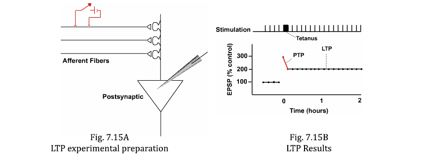
An enduring form of synaptic plasticity called long-term potentiation (LTP) is believed to be involved in many examples of declarative memory. It is present in the hippocampus, which is known to be involved in declarative memories. LTP can be studied in brain slice preparations where an electric shock (test stimulus) can be delivered to afferent fibers and the resultant summated EPSP can be recorded in the postsynaptic neuron (Figure 7.15A). If the pathway is repeatedly stimulated (e.g., every minute), the amplitude of EPSP is constant (Figure 7.15B).
Delivering a brief 1-sec duration train of high frequency (100 Hz) stimuli (i.e., the tetanus) to the afferent nerve produces two types of enhancement in the postsynaptic neuron. First, there is a transient facilitation called post-tetanic potentiation (PTP) that dies away after several minutes. Second, following the PTP is a very enduring enhancement of the EPSP called LTP. LTP is the kind of mechanism necessary to store a long-term memory (Figure 7.15B).
Figure 7.16
Animation of the induction and expression of LTP.The NMDA-type glutamate receptor is critical for some forms of LTP, in particular LTP at the CA3-CA1 synapse in the hippocampus. The postsynaptic spines of CA1 neurons have two types of glutamate receptors; NMDA-type glutamate receptors and AMPA-type glutamate receptors (Figures 7.16A). Both receptors are permeable to Na+ and K+, but the NMDA-type has two additional features. First, in addition to being permeable to Na+, it also has a significant permeability to Ca2+. Second, this channel is normally blocked by Mg2+.
Even if glutamate binds to the NMDA receptor and produces a conformational change, there is no efflux of K+ or influx of Na+ and Ca2+ because the channel is "plugged up" or blocked by the Mg2+. Thus, a weak test stimulus will not open this channel because it is blocked by Mg2+. A weak test stimulus will produce an EPSP, but that EPSP will be mediated by the AMPA receptor. It is as if the NMDA receptor were not even there.
Now consider the consequences of delivering a tetanus (Figure 7.16B). During the tetanus, there will be spatial and temporal summation of the EPSPs produced by the multiple afferent synapses on the common postsynaptic cell (Figure 7.15A). Consequently, the membrane potential of the postsynaptic neuron will be depolarized significantly, much more so than the depolarization produced by a single afferent test stimulus. Because the inside of the cell becomes positive with the large synaptic input, the positively charged Mg2+ is repelled by the inside positivity and is "thrust" out of the channel. Now the channel is unplugged and Ca2+ can enter the spine through the unblocked NMDA receptor. The Ca2+ that enters the cell activates various protein kinases, which then trigger long-term changes. One component of the long-term change is the insertion of new AMPA receptors into the postsynaptic membrane (Figure 7.16C). Therefore, after the tetanus, the transmitter released from the presynaptic neuron by a test stimulus will bind to a greater number of receptors on the postsynaptic neuron. If more receptors are bound and hence opened, a larger (potentiated) EPSP (i.e., LTP) will be produced (Figure 7.16C). In addition to an increase in the number of postsynaptic AMPA receptors, there is evidence that a greater amount of transmitter is released from the presynaptic neurons. The combination of the presynaptic and postsynaptic effects would act synergistically to increase the size of the synaptic potential in the postsynaptic neuron. Note that this example of a synaptic mechanism for declarative memory bears some similarity to the synaptic mechanism for the example of nondeclarative memory (sensitization) discussed previously. Although the specific details differ, both involve activation of second messenger systems and regulation of membrane channels. Therefore, at a fundamental mechanistic level, there does not appear to be significant differences between the two major classes of memory systems. The major difference appears to be the brain region and the neural circuit and into which the learning mechanism is embedded.
7.5 Enhancing Memory
With a knowledge of some of the genes and proteins involved in memory, we can use this information to try to both test the role of specific proteins in memory and also to improve memory. One experimental way of approaching the issue is to use transgenic technology in which a gene of interest can be over expressed in an animal by introducing it into an egg cell. When the offspring develop into adults, their performance on memory tests can be examined. An example of this approach is illustrated in Figure 7.17. Here the role of the NMDA receptor was examined by Joe Tsien and his colleagues, who were then at Princeton University. If NMDA receptors are important for the induction of LTP, and LTP is important for declarative memory, one would expect that animals that had a greater number of NMDA receptors would learn more readily. NMDA receptors were over expressed in mice and the mice were tested on the object discrimination test that was discussed earlier in the Chapter.
To assess the performance of a mouse on the object recognition task, the experimenter measures the amount of time for some predefined period the mouse spends exploring the one object, versus the amount of time the mouse spends exploring the other object. If the mouse remembers that it had seen one of the objects previously, it will spend more time exploring the novel one. As illustrated in Figure 7.17, one hour after the initial presentation of the objects, the mice do very well on the test. Indeed, they are correct about 100% of the time. They know the novel object. However, one day later the memory performance is rather poor, and after three days it is even worse. By one week, mice show no recognition memory.
What about the mice that received the extra NMDA receptors? Now one day after training they have perfect memory! So the extra receptors have led to an improved memory performance. That’s the good news – but the bad news is that the memory is no better one week later. This somewhat disappointing finding should not be surprising. Although NMDA receptors are important in memory, they are not the whole story. As indicated earlier in the Chapter, memory involves the synergistic engagement of multiple genes and proteins. So to improve memory further, it will be necessary to manipulate multiple genes. At the present time it is difficult to do so, but, it probably will become possible in the near future. It will also be possible to over express genes of interest in targeted areas of the human brain. The future for treating individuals with memory disabilities looks very promising.
This animation by Graduate students Julia Hill and Natalia Rozas De O'Laughlin of the Neuroscience Graduate Program at McGovern Medical School at UTHealth explains the concept of synaptic plasticity. It placed third in the 2011 Inaugural Society for Neuroscience Brain Awareness Video Contest.
- Question 1
- A
- B
- C
- D
- E
A 50-year old patient with recent damage to the hippocampus from a stroke would likely have all of the following deficits EXCEPT:
A. Difficulty learning new facts
B. Difficulty describing a recent event
C. Difficulty learning a new vocabulary word
D. Difficulty recalling a childhood memory
E. Difficulty remembering a face
A 50-year old patient with recent damage to the hippocampus from a stroke would likely have all of the following deficits EXCEPT:
A. Difficulty learning new facts This answer is INCORRECT.
The hippocampus is involved in declarative memory including the memory for facts.
B. Difficulty describing a recent event
C. Difficulty learning a new vocabulary word
D. Difficulty recalling a childhood memory
E. Difficulty remembering a face
A 50-year old patient with recent damage to the hippocampus from a stroke would likely have all of the following deficits EXCEPT:
A. Difficulty learning new facts
B. Difficulty describing a recent event This answer is INCORRECT.
The hippocampus is involved in declarative memory including the memory for recent events.
C. Difficulty learning a new vocabulary word
D. Difficulty recalling a childhood memory
E. Difficulty remembering a face
A 50-year old patient with recent damage to the hippocampus from a stroke would likely have all of the following deficits EXCEPT:
A. Difficulty learning new facts
B. Difficulty describing a recent event
C. Difficulty learning a new vocabulary word This answer is INCORRECT.
The hippocampus is involved in declarative memory including the memory for vocabulary words (semantic memory).
D. Difficulty recalling a childhood memory
E. Difficulty remembering a face
A 50-year old patient with recent damage to the hippocampus from a stroke would likely have all of the following deficits EXCEPT:
A. Difficulty learning new facts
B. Difficulty describing a recent event
C. Difficulty learning a new vocabulary word
D. Difficulty recalling a childhood memory This answer is CORRECT!
The hippocampus is involved in the formation of new memories, but not in the storage of old memories after they have been consolidated.
E. Difficulty remembering a face
A 50-year old patient with recent damage to the hippocampus from a stroke would likely have all of the following deficits EXCEPT:
A. Difficulty learning new facts
B. Difficulty describing a recent event
C. Difficulty learning a new vocabulary word
D. Difficulty recalling a childhood memory
E. Difficulty remembering a face This answer is INCORRECT.
The hippocampus is involved in object recognition.
- Question 2
- A
- B
- C
- D
Short term memories can involve all of the following processes EXCEPT:
A. Regulation of gene expression
B. Activation of second-messenger systems
C. Modulation of membrane channels
D. Modulation of transmitter release
Short term memories can involve all of the following processes EXCEPT:
A. Regulation of gene expression This answer is CORRECT!
Regulation of gene expression is associated with long-term memories and not short-term memories.
B. Activation of second-messenger systems
C. Modulation of membrane channels
D. Modulation of transmitter release
Short term memories can involve all of the following processes EXCEPT:
A. Regulation of gene expression
B. Activation of second-messenger systems This answer is INCORRECT.
Activation of second-messenger systems such as cAMP is associated with short-term memory.
C. Modulation of membrane channels
D. Modulation of transmitter release
Short term memories can involve all of the following processes EXCEPT:
A. Regulation of gene expression
B. Activation of second-messenger systems
C. Modulation of membrane channels This answer is INCORRECT.
Both voltage-gated and transmitter-gated channels are associated with short-term memory.
D. Modulation of transmitter release
Short term memories can involve all of the following processes EXCEPT:
A. Regulation of gene expression
B. Activation of second-messenger systems
C. Modulation of membrane channels
D. Modulation of transmitter release This answer is INCORRECT.
Changes in synaptic strength are associated with short-term memory.
- Question 3
- A
- B
- C
- D
- E
Classical conditioning is an example of:
A. Semantic memory
B. Episodic memory
C. Implicit memory
D. Declarative memory
E. Nonassociative memory
Classical conditioning is an example of:
A. Semantic memory This answer is INCORRECT.
Semantic memory is a type of declarative memory, whereas classical conditioning is a type of nondeclarative (implicit) memory.
B. Episodic memory
C. Implicit memory
D. Declarative memory
E. Nonassociative memory
Classical conditioning is an example of:
A. Semantic memory
B. Episodic memory This answer is INCORRECT.
Episodic memory is a type of declarative memory, whereas classical conditioning is a type of nondeclarative (implicit) memory.
C. Implicit memory
D. Declarative memory
E. Nonassociative memory
Classical conditioning is an example of:
A. Semantic memory
B. Episodic memory
C. Implicit memory This answer is CORRECT!
D. Declarative memory
E. Nonassociative memory
Classical conditioning is an example of:
A. Semantic memory
B. Episodic memory
C. Implicit memory
D. Declarative memory This answer is INCORRECT.
Classical conditioning is an example of nondeclarative memory.
E. Nonassociative memory
Classical conditioning is an example of:
A. Semantic memory
B. Episodic memory
C. Implicit memory
D. Declarative memory
E. Nonassociative memory This answer is INCORRECT.
Classical conditioning is a form of associative learning, which is in contrast to examples of nonassociative memory like sensitization.