Lab 2 (ƒ4) - External and Internal Anatomy of the Spinal Cord
External Landmarks - External Diagram
 Look at the diagram of the spinal cord and the gross specimen provided in lab.
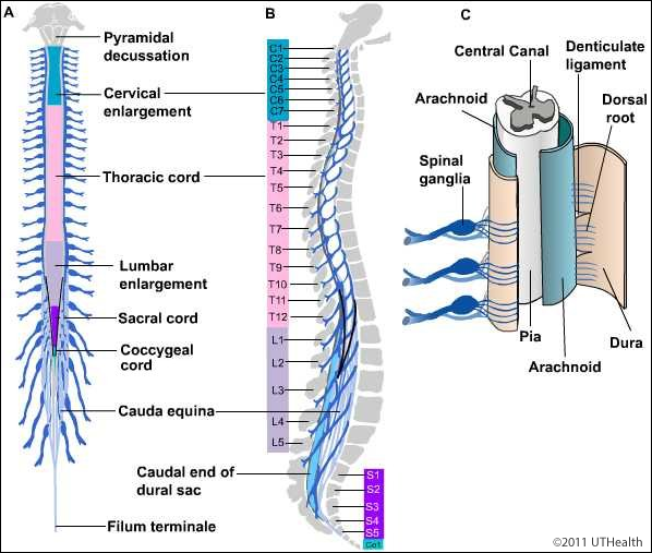
Nerve rootlets emerge from the posterolateral and anterolateral parts of the cord along its entire length. You will notice that a few centimeters from the cord, groups of nerve rootlets fuse together to form the posterior and anterior roots (normally 31 pairs in the intact spinal cord).
Each pair of roots passes through one of the intervertebral spaces of the spinal column. The spinal cord consists of 31 segments defined by their corresponding pair of posterior and anterior roots (8 cervical, 12 thoracic, 5 lumbar, 5 sacral, and 1 coccygeal).