Lab 11 - The Limbic System
Hypothalamus - Introduction
The purpose of this section is to identify and learn the:
- Regions of the hypothalamus.
- Nuclei within regions of the hypothalamus.
- Main pathways which interconnect the hypothalamus with other brain regions.
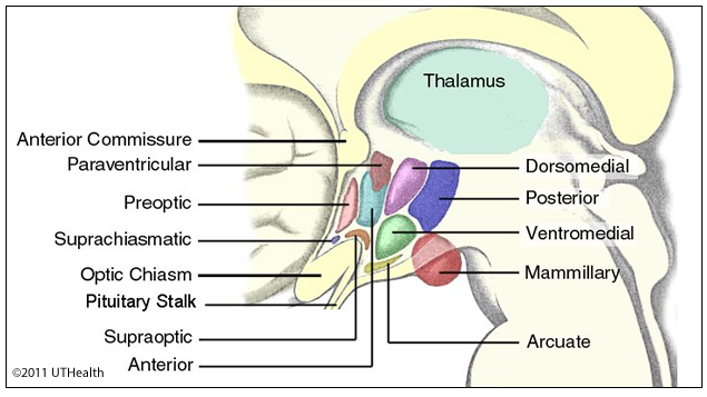 On the medial surface of the hemisected brain there is a shallow depression (hypothalamic sulcus) that separates the hypothalamus from the thalamus. The part of the hypothalamus, which you can see, is the medial surface of a thin wall, which surrounds the third ventricle. This wall, which contains hypothalamic nuclei and fibers passing through, extends laterally only a few millimeters beyond the midline. Locate the mammillary bodies, the infundibulum (hypophyseal stalk), and the tuber cinereum, which surrounds the upper neck of the infundibulum. A thin membrane called the lamina terminalis marks the rostral limit of the hypothalamus and the third ventricle.
The hypothalamus contains three major regions that extend caudally from the lamina terminalis: (1) anterior region, directly above the optic chiasm and is bordered dorsally by the hypothalamic sulcus; (2) tuberal region, includes the tuber cinereum and the area above which extends to the hypothalamic sulcus, and (3) posterior (mammillary) region, includes the mammillary bodies and the area above which extends to the hypothalamic sulcus.
Use the indicated landmarks to identify the hypothalamic regions on the medial surface of the hemisected brain.