Lab 10 (ƒ2) - Internal Organization of the Brain
Internal Capsule
The internal capsule is composed of all of the fibers going to and coming from the cerebral cortex which form a fan-shaped mass of fibers called the corona radiata. The fibers of the corona radiata converge towards the diencephalon forming a prominent, compact bundle of axons known as the internal capsule which links the cortex to other levels of the neuraxis.
Fibers within the internal capsule connect the thalamus with the cerebral cortex by thalamocortical and corticothalamic fibers. In addition to these fibers, the internal capsule contains axons which descend from the cortex to lower levels of the neuraxis such as the brain stem and spinal cord. These cortical efferent fiber systems, which are referred to as corticofugal fibers, include the corticoreticular, corticopontine, corticobulbar and corticospinal tracts. Smaller projections also exist to the basal ganglia, other areas of the diencephalon and the midbrain.
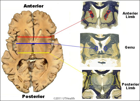
The Internal Capsule can be divided into three parts. These parts (anterior limb, genu, and posterior limb) are best appreciated in horizontal sections. The anterior limb is positioned between the lenticular nucleus (comprised of the putamen and globus pallidus) and the head of the caudate (we will learn about these structures later). The Posterior Limb is positioned between the lenticular nucleus and the thalamus. The Genu (or knee) is the portion between the anterior and posterior limbs, and is the section closest to the midline.